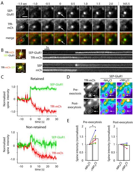
Figure 3. AMPA Receptors are Directly Inserted Into the Spine Plasma Membrane.
(A) Time lapse imaging of a hippocampal neuron coexpressing SEP-GluR1 (top row) and TfR-mCh (middle row) following stimulation with Bic/Gly. Note the abrupt appearance of SEP-GluR1 fluorescence at the precise location of a spine RE (arrows). Scale bar, 1 μm.
(B) Kymograph comparison of newly inserted synaptic cargo. Shown are single exocytic events of SEP-GluR1 (top) and TfR-mCh-SEP (bottom). Note that SEP-GluR1 is inserted and retained in the spine head (upper kymograph), even as TfR-mCh from the same endosome quickly diffuses away with similar kinetics as newly exocytosed TfR-mCh-SEP (lower kymographs). Scale bar, 0.5 μm.
(C) SEP-GluR1 exocytic events fall into two classes. Following RE fusion, SEP-GluR1 was either retained in spines (62% of events, top panel) or quickly diffused away (38% of events, bottom panel). In all cases, colocalized TfR-mCh signal (red traces) declined rapidly following the exocytosis of SEP-GluR1 (green traces).
(D) Exocytosis of SEP-GluR1 occurs in an all-or-none manner. Prior to stimulation, cells were treated with 50 mM NH4Cl to reveal SEP-GluR1 within spine REs. Following RE exocytosis, NH4Cl had no effect on SEP-GluR1 intensity in the same spines indicating plasma membrane localization. Scale bar, 1 μm.
(E) Normalized spine SEP-GluR1 fluorescence intensity with and without NH4Cl before and after SEP-GluR1 exocytosis in the same spines (* p<0.05, paired students t-test). See also Movie S3.

