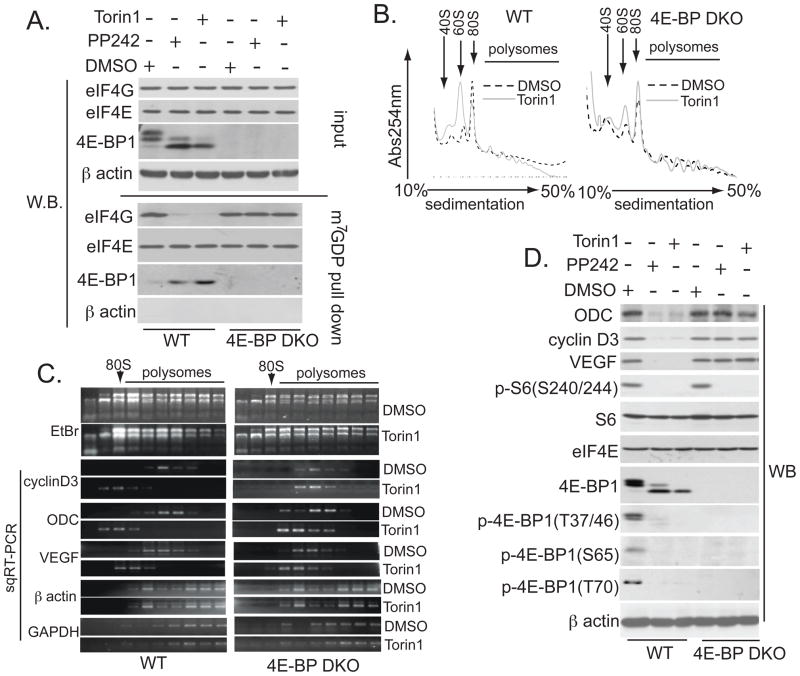
Fig. 4. asTORi inhibit translation of mRNAs encoding proliferation-promoting proteins via 4E-BPs.
(A) WT and 4E-BP DKO MEFs treated for 24 hours with 2.5 μM PP242, 250 nM Torin1, or vehicle (DMSO) were subjected to m7GDP pull-down. Amounts of the indicated proteins in the input (10%) or pull-down (25%) were determined by Western blotting. β actin served as a loading control (input) and to exclude contamination (m7GDP pull-down). (B) Absorption profiles of ribosomes from WT and 4E-BP DKO cells treated for 24 hours with 250 nM Torin1, or vehicle (DMSO). 40S and 60S denote the corresponding ribosomal subunits; 80S, monosome. (C) RNA was visualized by ethidium bromide (EtBr). Distribution of cyclin D3, VEGF, ODC, β actin, and GAPDH mRNAs was determined by semiquantitative reverse transcription polymerase chain reaction (sqRT-PCR). The RT-PCR reactions were in the linear range (fig. S7I). (D) WT and 4E-BP DKO MEFs were treated as in (A), and the amount of the indicated proteins was determined by Western blotting. β actin served as a loading control.

