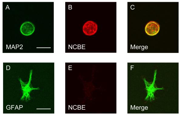
Fig. 10.
Expression of NCBE in freshly dissociated mouse hippocampal neurons. (A-C) Immunocytochemistry of a freshly dissociated HC neurons doubled stained with MAP2 (green) and NCBE antibody (red). (D-F) Immunohistochemistry of a freshly dissociated HC astrocyte double-stained with GFAP (green) and NCBE antibody (red). The results are representative of three experiments. Scale bar: 10 μm.

