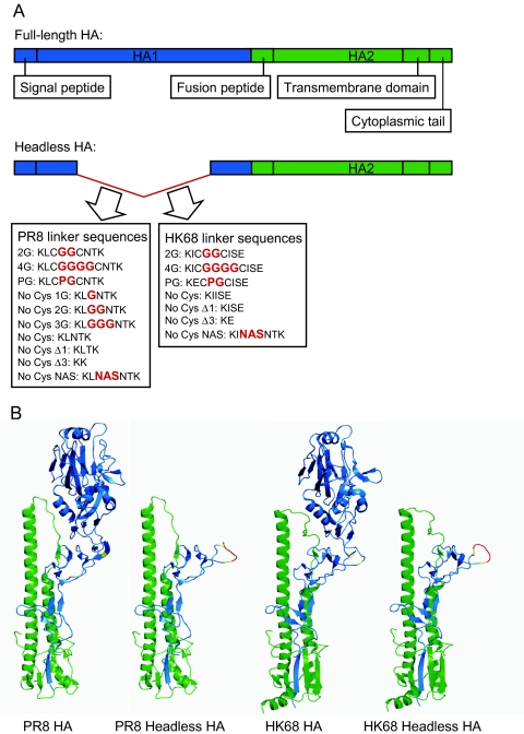
FIG 1 .
Schematic of headless HA constructs. (A) Schematic of the linear structure of the full-length influenza virus HA protein (top) and a generalized headless HA protein (bottom). Linker peptides tested in the context of the PR8 and HK68 HA sequences are shown. Inserted amino acids are shown in bold red type, while amino acids present in the native HA sequence are in black. (B) Schematic of the folded structures of the full-length and headless HAs of PR8 virus (left panel) and HK68 virus (right panel). In both cases, headless HAs carrying the 4G linker bridge are depicted. The HA1 subunit is blue, and the HA2 subunit is green. Cysteines 52 and 277 are shown in yellow, and 4G linker sequences are shown in red. The full-length HA structures were downloaded from the Protein Database (PDB): PR8 HA (PDB ID 1RVX) and HK68 HA (PDB ID 1MQN). Schematics of headless HAs were generated using the full-length HA coordinates as a starting point, and 4G loops were manually docked into the headless HA carbon to close the discontinuous alpha-carbon amino acid chain. Final images were generated with PyMol (Delano Scientific).

