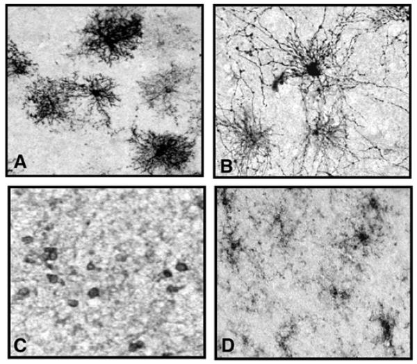
Fig. (2).
Photomicrographs displaying the different types of immunopositive neuroglia in the prefrontal cortex of a psychiatrically normal human subject. Images A and B show two types of astroglia both immunostained with an anti-GFAP (glial fibrillary acid protein) antibody which is a marker for reactive astroglia. Protoplasmic astrocytes (A) are found mainly in cortical gray matter, and fibrous astrocytes (B) are found mainly in cortical white matter. Image C displays oligodendrocytes in cortical white matter which are immunostained with anti CNPase antibody, a marker for mature oligodendrocytes. Image D illustrates microglia in white matter immunostained for anti LFA-1 antibody, a marker of active microglia. All photomicrographs were taken with the 20x objective of a Nikon Eclipse E600 microscope.

