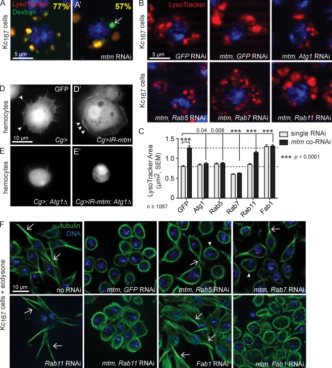
Figure 5.
mtm interacts with both endocytic and autophagic effectors, with separable effects on endolysosomes and cell remodeling. (A and A’) Arrow indicates delay in late F-dextran (green) trafficking to LysoTracker-positive organelles (red) in mtm RNAi Kc167 cells, shown as the percent of colocalization 20 min after uptake. (B and C) Screen of endolysosome size in single and mtm coRNAi–treated Kc167 cells, identified suppressors of mtm-enlarged endolysosomes. (B) LysoTracker (red) with coRNAi as shown. (C) Quantification of mean endolysosomal area upon single RNAi (gray) or mtm coRNAi (black). Bottom dashed line, mean area GFP RNAi; top dashed line, mean area of mtm, GFP coRNAi. (D) GFP-labeled Atg1Δ3D/+ hemocytes with extended protrusions (arrowheads). (D’) Lack of cell protrusions (ruffles, arrowheads) in spread mtm-depleted hemocytes. (E) Round Atg1Δ3D−/− hemocytes failed to spread. (E’) Round cells with Atg1Δ3D−/− in combination with mtm depletion. (F) Microtubules (green) in Kc167 cells 1 d after ecdysone with coRNAi as shown. With mtm and Rab5 or Rab7 codepletion, mixed examples of cells that partially reverted cell extensions (arrows) and cells that remained round (arrowheads). No effect of Rab11 or Fab1 RNAi alone or on mtm RNAi defect. Error bars indicate SEM.

