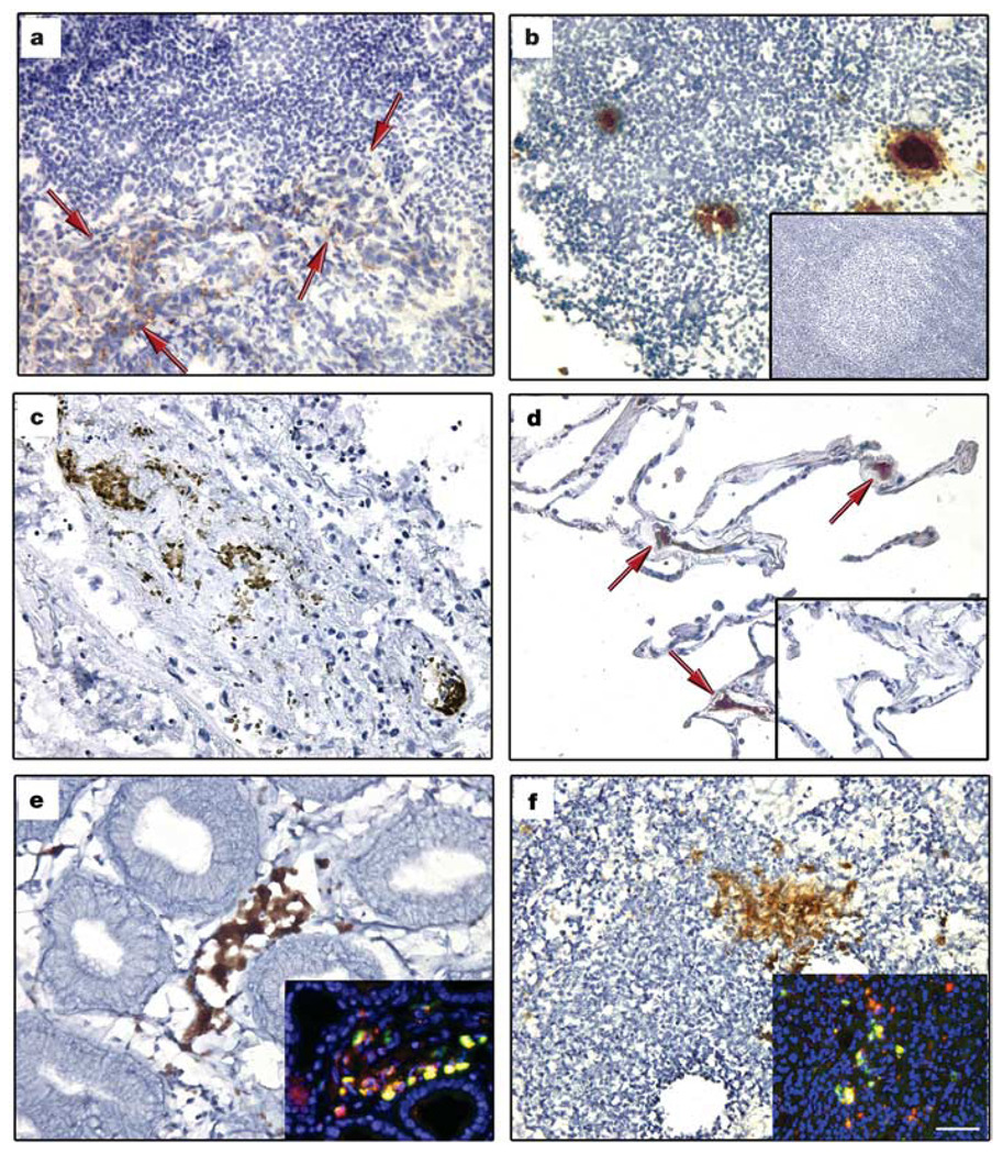
Figure 3. Expression of VEGFR1 in pre-metastatic human tissue.
a–f, Cellular clusters stained with VEGFR1 in malignant and non-malignant tissues in individuals with breast (n = 15), lung (n = 15) and gastrointestinal (n = 3) cancers. Lymph node with evidence of breast adenocarcinoma metastasis (a, red arrows indicate tumour) and lymph node without malignancy from same patient (b). Primary lung adenocarcinoma (c) and adjacent ‘normal’ lung without neoplasm (d, red arrows indicate VEGFR1+ cells). No VEGFR1+ clusters were seen in lymph node (b, inset; n = 6) and lung tissue (d, inset; n = 3) from individuals without cancer. Also shown is a primary adenosquamous carcinoma of the gastrooesophageal junction (e), and a hepatic lymph node without carcinoma (f). Insets in e, f, show co-immunofluorescence of VEGFR1 (red) and c-Kit (green). Scale bar at bottom right applies to all panels (40 µm; insets, 40 µm).

