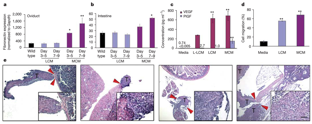
Figure 6. Redirection of LLC metastases to atypical sites.
a, b, By quantitative RT–PCR analysis, increased fibronectin expression was seen in the oviduct (a) and intestine (b) in mice given MCM compared with wild-type and LCM-treatment. For oviduct, *P < 0.05 at days 3–5 and **P < 0.001 for days 7–9 compared with wild type, and for intestine, *P < 0.001 at days 7–9 compared with wild type by ANOVA (n = 6). c, ELISA assay (in triplicate) for VEGF and PlGF levels in the conditioned media (*P < 0.05 when compared with L-LCM, **P < 0.01 when compared with media alone, by ANOVA). d, Transwell migration assays (in triplicate) demonstrate enhanced migration of VEGFR1+ cells to LCM and MCM (**P < 0.001 by ANOVA). e, Treatment with MCM redirects the metastatic spread of LLC to B16 melanoma metastatic sites, such as the spleen (left panel), kidney (left middle panel), intestine (right middle panel) and oviduct (right panel). Arrows denote the regions of metastatic borders, which are shown in the insets (n = 6). T, LLC tumour cells. Scale bar at bottom right applies to panel e (200 µm; insets, 20 µm).

