
Figure 1. Altered connectivity in children with 22q11.2 deletion syndrome.
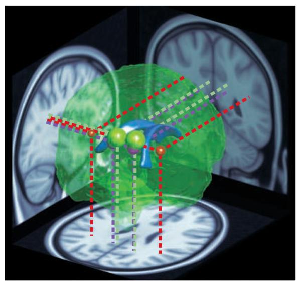
A reconstruction of the brain viewed from the posterior right side. The lateral ventricles (shown in blue) are depicted in the centre as a point of reference. The coloured spheres indicate the location and approximate extent of major clusters of reduced fractional anisotropy (FA) — a measure of neural connectivity — in children with 22q11.2 deletion syndrome (22q11.2DS), as reported by REF. 32 (red), REF. 36 (green) and REF. 87 (purple). The data were obtained using diffusion tensor imaging, an MRI technique that measures the directionality of water diffusion in the brain as an indicator of the organization and integrity of neuronal tracts wrapped in myelin (white matter). The location and size of the coloured spheres depict the sites of peak FA differences and their extent. Projections onto brain slices (dotted lines) indicate the positions of the clusters in major white matter tracts. The red spheres represent the left and right extremities in a wedge-shaped cluster reported by REF. 32. The entire area between them showed higher FA in children with 22q11.2DS compared with typical developing controls, but because this large cluster would completely envelope the other regions, it is not represented here in its entirety. In summary, three separate studies from two different teams have reported similar, and in one case directly overlapping, differences in neural connectivity in children and young adults with 22q11.2DS.