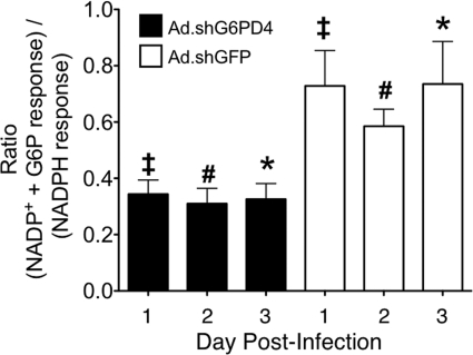
FIGURE 6.
shRNA-G6PD-mediated knockdown in G6PD-deficient mice further reduces nuclear superoxide production in response to NADP+ + G6P. G6PD homozygous-deficient (MT) mice were infected with Ad.shG6PD4 (black bars) or with Ad.shGFP (white bars). The liver nuclei were isolated 1, 2, or 3 days after infection. The nuclei were treated with NADP+ + G6P or with NADPH. EPR was performed, and the ratios of [DMPO-OH] signal with each substrate are plotted as described for Fig. 4F. The results depict the mean ± S.E. for n = 3–5 mice for each experimental point. Statistical significance was tested using ANOVA and Bonferroni's multiple comparison test. The symbols indicating statistical significance (p < 0.05) are as follows: ‡, day 1 Ad.shG6PD4 versus day 1 Ad.shGFP. #, day 2 Ad.shG6PD4 versus day 2 Ad.shGFP. *, day 3 Ad.shG6PD4 versus day 3 Ad.shGFP.

