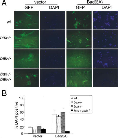
Figure 2.
BAD(3A) does not induce apoptosis in bax−/−bak−/− MEF. (A) MEF of different genotypes were infected with GFP (vector) or BAD(3A)–IRES–GFP [Bad(3A)]. Twenty-four hours after infection, cells were stained with DAPI, and photographed by use of a FITC or DAPI filter. (B) Cells from A were collected and subjected to FACS analysis. The percentage of dead cells was determined by the ratio of DAPI-positive cells to GFP-positive cells. Data shown is the average of three independent experiments.

