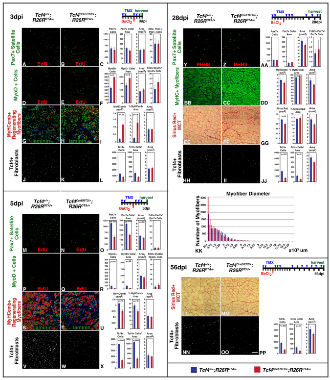
Fig. 6.
During muscle regeneration, ablation of Tcf4+ fibroblasts leads to premature satellite cell differentiation and smaller regenerated myofibers. (A-L) At 3 dpi, Tcf4+ cells are reduced (J-L), with no change in Pax7+ cells (A-C), but with an increase in MyoD+ progenitors/myoblasts (D-F) and MyHCemb+ regenerating myofibers (G-I) in Tcf4CreERT2/+;R26RDTA/+ mice. (M-X) At 5 dpi, 42% of Tcf4+ cells were calculated to be ablated (V-X), resulting in fewer Pax7+ cells (M-O), MyoD+ progenitors/myoblasts (P-R) and MyHCemb+ regenerating myofibers (S-U) in Tcf4CreERT2/+;R26RDTA/+ mice. (Y-KK) At 28 dpi, despite Tcf4+ fibroblast ablation (HH-JJ), Pax7+ cells recover (Y-AA), muscle largely regenerates (BB-GG), but diameter of myofibers is reduced (BB,CC,EE,FF,KK) in Tcf4CreERT2/+;R26RDTA/+ versus Tcf4+/+;R26RDTA/+ mice. (LL-PP) By 56 dpi, regenerated muscle is recovered in Tcf4CreERT2/+;R26RDTA/+ mice. Scale bars: in OO,100 μm for all photomicrographs. For all graphs, mean ± s.e.m. are plotted.

