
Figure 2.
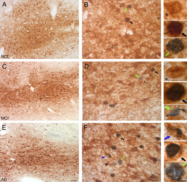
p75NTR/pS422 Immunolabeled neurons. Sections containing the NB subfield of the CBF dual immunostained for p75NTR+ (brown) and pS422+ (blue) show a decrease in the number of p75NTR+ neurons with an increase of pS422+ neurons as disease progression continues from NCI (A and B), to MCI (C and D) and AD (E and F). In NCI (B), most NB neurons were single-labeled for p75NTR+ (asterisk), few neurons were double-labeled for p75NTR+/pS422+ (black arrow), whereas only an occasional neuron was single-labeled for the early tau pathological marker, pS422+ (green arrow). In the MCI (D) and AD (F) cases, there was a decrease in the p75NTR+ neurons (asterisk) and an increase in both p75NTR+/pS422+ (black arrow) and pS422+ (green arrow) neurons. In some p75NTR+/pS422+ neurons, it can be seen that the pS422 immunoreactivity begins in the center of the neuron (blue arrow). Images in A, C, and E were taken at magnification ×4. Scale bar = 250 μm. Images in B, D, and F were taken at magnification×20. Scale bar is 50 μm. The panels to the right were taken at magnification ×60. Scale bar is 25 μm.