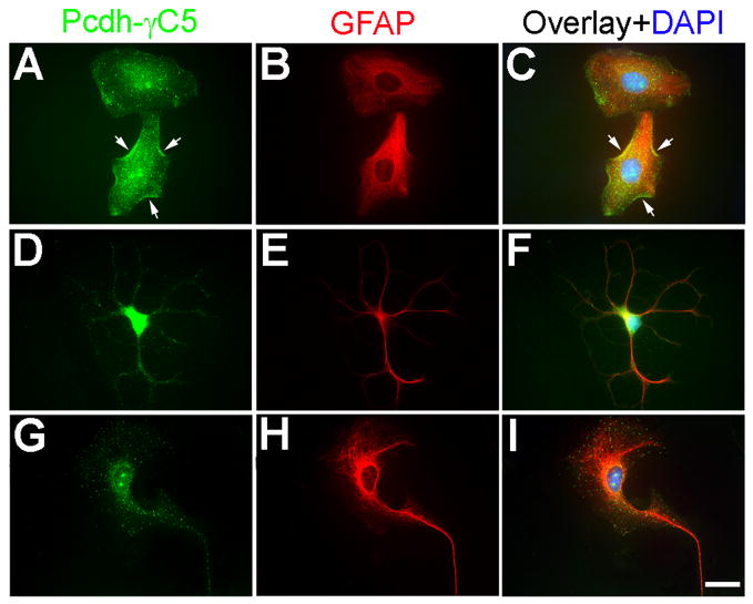
Fig. 6. Pcdh-γC5 is expressed by cultured astrocytes.
Triple-label fluorescence of cultured astrocytes with Rb anti-Pcdh-γC5 (green, A, D, G), mouse anti-GFAP (red, B, E, H) and the nuclear stain DAPI (blue, C, F, I). The Pcdh-γC5 is expressed in astrocytes with various morphologies. The Pcdh-γC5 immunofluorescence is frequently shown in the form of clusters both in the cell body and processes. Some of the clusters are clearly associated with the plasma membrane (A, C, arrows). Scale bar=20μm.

