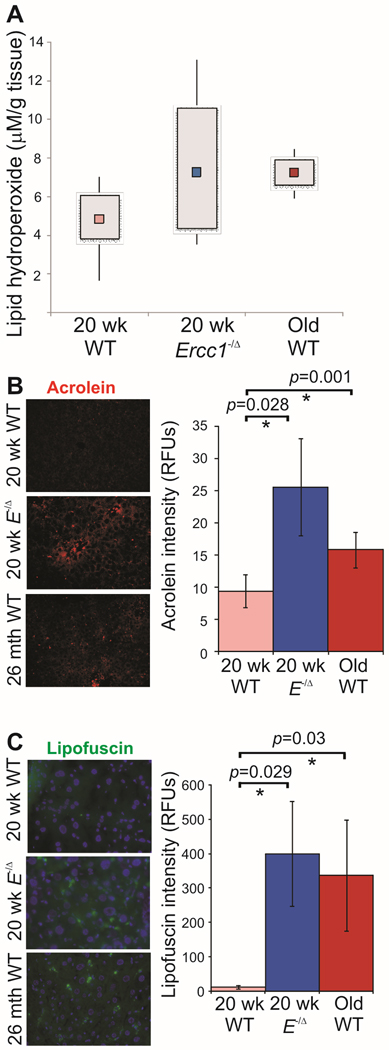
Figure 6. Increased oxidative damage in liver of old and progeroid mice.
(A) Immunodetection of lipid hydroperoxides, a by-product of lipid peroxidation, in liver lysates from 20 wk-old WT and Ercc1−/Δ littermate mice vs. old WT mice (26–34 months of age). The box plot represents the mean (in colored square), along with the 25th and 75th percentile for each group (n=5 mice). Average lipid hydroperoxide levels for groups were as follows: 20 wk WT (4.8µM/g liver), 20 wk Ercc1−/Δ (7.3µM/g liver; p=0.12, one-tailed Student’s t-test), and old WT (7.2µM/g liver).
(B) Immunofluorescence detection of acrolein, a by-product of lipid peroxidation. The staining intensity was quantified using AxioVision imaging software. Plotted are the averages ± S.E.M. determined from analysis of 10 random fields of liver sections from 2 mice per group. Asterisks indicate significant differences.
(C) Representative images illustrating lipofuscin accumulation in 20 week-old WT and Ercc1−/Δ mice and 26–34 month-old WT mouse liver. AxioVision software was used for quantification of 10 random fields measured from 5 mice per group. Plotted is the average fluorescence intensity for each group ± S.E.M. Asterisks indicate significant differences.

