
Figure 1.
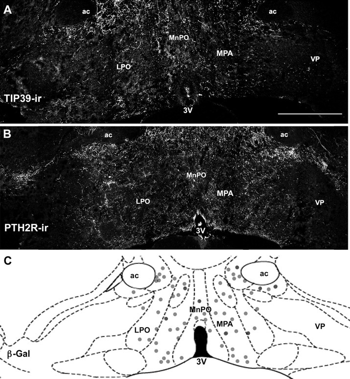
Expression of TIP39 and PTH2R immunoreactivity in the preoptic hypothalamus. A, B, Low-magnification images show TIP39-ir (A) and PTH2R-ir (B) concentrated in hypothalamic preoptic areas. C, Schematic representation of the preoptic hypothalamus at the same level shows the location of β-gal-expressing cells in a PTH2R-β-gal knock-in mouse. Scale bar (in A) A, B, 500 μm. 3V, Third ventricle; ac, anterior commissure; LPO, lateral preoptic area; VP, ventral pallidus.