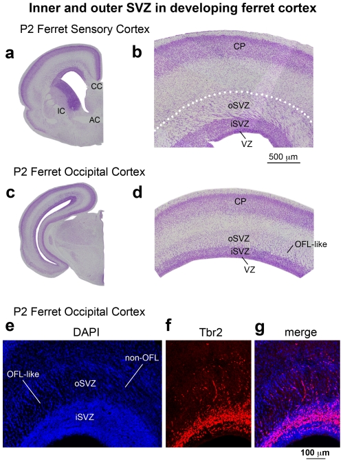
Figure 21. Inner and outer SVZ in the developing ferret cortex.
(a,c) Nissl-stained coronal sections of somatosensory and visual cortex from P2 ferret. (b,d) Higher magnification images taken from the sections shown in (a) and (c). Dotted line in (b) represents the outer boundary of the outer subventricular zone (oSVZ) in somatosensory cortex determined through Tbr2 immunostaining. The outer fiber layer (OFL) and inner fiber layer are not present in somatosensory cortex, but a structure that resembles the OFL (OFL-like) is apparent in some areas of the ferret occipital lobe. However, note that the OFL-like structure in ferret is located within the oSVZ, in contrast to the OFL in macaque, which is superficial to the oSVZ. In both somatosensory and visual cortex the boundary between the inner SVZ (iSVZ) and oSVZ can be visualized in Nissl stained tissue as a sharp border created by different cellular density. The superficial boundary of the oSVZ can also be visualized in Nissl stained tissue based on cell density (determined through Tbr2 immunostaining). In somatosensory cortex the oSVZ is characterized by a striated appearance of cells organized into clusters that appear to stream at an oblique or tangential angle through the oSVZ. (e–g) The OFL-like structure in ferret resembles the macaque OFL since radially oriented clusters of cells stream through this structure as they do in macaque (see Fig. 4). But the ferret OFL-like structure differs from the macaque OFL since it is within the oSVZ rather than superficial to the oSVZ and because Tbr2+ cells are located throughout the OFL-like structure in ferret but do not penetrate the macaque OFL (See Fig. 4). VZ, ventricular zone; CP, cortical plate. Scale bar in (b) applied to (d). Scale bar in (g) applied to (e, f).

