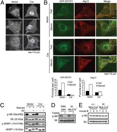
Fig. 3.
Localized production of PI(3)P, autophagosome formation, and mTOR signaling are impaired in the absence of Vps34. (A) Ablation of Vps34 leads to diminished PI(3)P production. Vps34f/f MEFs stably expressing GFP-FYVE were infected with vector control or Cre. Three representative cells from each group are shown. Note the lack of FYVE puncta in Cre-infected MEFs, indicating the absence of PI(3)P. (B) Serum starvation fails to induce DFCP1 and Atg12 puncta in Vps34-null cells. Vps34f/f MEFs were infected with vector control or Cre virus for 5 d. Cells were transiently transfected with GFP-DFCP1. Forty-eight hours after transfection, cells were fixed and immunofluorescence for Atg12 was performed. Representative images are shown. The average numbers of yellow or red puncta were determined from eight countings and are shown with SEM. Note that upon serum withdrawal, vector control cells exhibit colocalization of DFCP1 and Atg12 puncta. In contrast, no puncta formation was observed for either DFCP1 or Atg12 in the Vps34-null cells. (C) Amino acid-stimulated mTOR activation is compromised in the absence of Vps34. Vps34f/f MEFs infected with vector or Cre were starved and stimulated for 30 min with 2× MEM amino acids. mTOR signaling was measured by the levels of phospho-S6 and phospho-4EBP1. (D and E) Steady-state mTOR signaling is not affected by the loss of Vps34. Whole liver lysates from wild-type and Vps34f/f;Alb-Cre+ mice (D) and lysates from cardiomyocytes isolated from wild-type and Vps34f/f;Mck-Cre+ mice (E) were probed for phospho-S6 and total S6.

