Abstract
Early studies suggested androgen receptor (AR) splice variants might contribute to the progression of prostate cancer (PCa) into castration resistance. However, the therapeutic strategy to target these AR splice variants still remains unresolved. Through tissue survey of tumors from the same patients before and after castration resistance, we found that the expression of AR3, a major AR splice variant that lacks the AR ligand-binding domain, was substantially increased after castration resistance development. The currently used antiandrogen, Casodex, showed little growth suppression in CWR22Rv1 cells. Importantly, we found that AR degradation enhancer ASC-J9 could degrade both full-length (fAR) and AR3 in CWR22Rv1 cells as well as in C4-2 and C81 cells with addition of AR3. The consequences of such degradation of both fAR and AR3 might then result in the inhibition of AR transcriptional activity and cell growth in vitro. More importantly, suppression of AR3 specifically by short-hairpin AR3 or degradation of AR3 by ASC-J9 resulted in suppression of AR transcriptional activity and cell growth in CWR22Rv1-fARKD (fAR knockdown) cells in which DHT failed to induce, suggesting the importance of targeting AR3. Finally, we demonstrated the in vivo therapeutic effects of ASC-J9 by showing the inhibition of PCa growth using the xenografted model of CWR22Rv1 cells orthotopically implanted into castrated nude mice with undetectable serum testosterone. These results suggested that targeting both fAR- and AR3-mediated PCa growth by ASC-J9 may represent the novel therapeutic approach to suppress castration-resistant PCa. Successful clinical trials targeting both fAR and AR3 may help us to battle castration-resistant PCa in the future.
Introduction
Prostate cancer (PCa) is currently the second leading cause of death in men in the United States [1]. Androgen deprivation therapy (ADT) has been the standard treatment for patients with advanced PCa since Huggins and Hodges [2] reported the castration effect on PCa. ADT is initially effective to inhibit the growth of androgen-dependent PCa and suppresses tumor progression in most PCa patients; however, most patients treated with current ADT eventually progress with castration-resistant PCa (CRPC) within 1 to 2 years [3,4]. The mechanisms underlying castration-resistant androgen receptor (AR)-mediated signaling remain unclear, although several possible mechanisms have been proposed [5–11].
One proposed mechanism involves the AR splice variants, especially AR3 (also named as AR-V7) that lacks the portion of the ligand-binding domain (LBD) [8,9], which have been reported to transactivate AR-targeted genes in the absence of androgen [7–10,12]. Interestingly, a recent report from Watson et al. [12] indicated that such constitutively active AR splice variants (AR-V7) might require full-length AR (fAR). They demonstrated that the growth of LNCaP cells with AR-V7 overexpression was suppressed after MDV3100 (a new antiandrogen) treatment or using small interfering RNA to target fAR. These findings raised an interesting question as to whether those AR splice variants have any translational or clinical value to target.
We report here that AR3 might represent an important target to suppress owing to its roles at selective stage(s) of PCa progression. Furthermore, we demonstrated that AR degradation enhancer, ASC-J9, was able to degrade both fAR and AR3 that resulted in the suppression of AR-targeted genes expression and cell growth in several CRPC cells.
Materials and Methods
Cells, Reagents, and Human Prostate Specimens
Human PCa cells CWR22Rv1, CWR22Rv1-fARKD (knockdown of fAR [13]), C4-2, and C81 were used. The antibodies for AR (N-20) and glyceraldehyde 3-phosphate dehydrogenase (GAPDH) were purchased from Santa Cruz Biotechnology (Santa Cruz, CA). The antibody for AR-V7 was kindly provided by Dr Jun Luo [8]. ASC-J9 (5-hydroxy-1,7-bis(3,4-dimethoxyphenyl)-1,4,6-heptatrien-3-one), also named as dimethylcurcumin, was a gift from AndroScience (San Diego, CA), and bicalutamide (Casodex) was purchased from AstraZeneca (Wilmington, DE). Plasmids containing AR3 complementary DNA and short hairpin RNAs specific for AR3 (shAR3) were kindly provided by Dr Yun Qiu [9]. Human primary prostate tissues were collected from the same patients before ADT and after development to CRPC at Tohoku University Hospital (Japan), Miyagi Cancer Center (Japan), and Chang Gung Memorial Hospital (Taiwan). These patients underwent transrectal prostate needle biopsy or transurethral resection of the prostate. This study has been approved by the ethics committee of the three institutions (Tohoku University Hospital, Miyagi Cancer Center, and Chang Gung Memorial Hospital), and informed consent was obtained from each patient. The patients' characteristics (age, prostate-specific antigen [PSA] level, Gleason score, stage, and time to develop CRPC) and outcomes (sample harvest after progression to CRPC, survival time after ADT, and the current status of alive or death) are summarized in Table W1.
Western Blot Analysis, Quantitative Real-time Polymerase Chain Reaction, and Luciferase Reporter Assay
Cells were cultured and treated with or without ASC-J9 for 24 hours in 10% charcoal-dextran-stripped fetal bovine serum (CD-FBS) media. Cell lysates were harvested and subjected to Western blot analysis. Quantitative real-time polymerase chain reaction (qPCR) was performed in triplicate with a Bio-Rad iCycler system (Bio-Rad, Hercules, CA); and messenger RNA (mRNA) levels of PSA, TMPRSS2, FKBP5, and GAPDH were measured. Cells were transiently transfected with mouse mammary tumor virus luciferase reporter (MMTV-Luc) or ARE4-Luc plus pRL-TK as internal control. Luciferase activities were measured using GloMax 20/20 Luminometer (Promega, Madison, WI).
Cell Growth Assay
Cells were treated with vehicle, 1 nM dihydrotestosterone (DHT), 5 µM Casodex, and 5 or 10 µM ASC-J9 in 10% CD-FBS medium. The media were replenished every other day, and we followed the standard MTT assay protocol.
Immunohistochemical Analysis
The paraffin-embedded tissue sections were stained with anti-ARV7 and counterstained with hematoxylin. These staining signals were manually evaluated by a pathologist (H.M.) blinded to patient identity. Each sample was classified with negative, weak, moderate, and strong expression based on intensity score and the percentage of immunoreactive cells [14].
In Vivo Tumor Growth Assay
Animal procedures were conducted in accordance with the protocol approved by the University of Rochester Committee on Animal Resources. CWR22Rv1 cells (1 x 10⁶ cells per site) were injected into both anterior prostates (orthotopic) of castrated nude mouse after 2 weeks of implantation. The mice were randomly divided into two groups (four mice/eight tumors each group) and either received 75 mg/kg ASC-J9 intraperitoneal injection or vehicle control every other day. After 4 weeks of treatment, all mice were killed to examine the tumor growth. Body weights and mice activity were measured weekly.
Statistical Analysis
All values are presented as mean ± SEM. Statistical analyses and comparisons among groups were performed using Student's t test (2-tailed). P < .05 was considered statistically significant.
Results
Casodex Has Little Effect on Growth of Castration-Resistant PCa Cell Lines
We first surveyed AR3 expression in various castration-resistant human PCa cell lines and found that the expression amount of AR3 (80 kDa) was very low in C81 and C4-2 cells but was abundant in CWR22Rv1 cells (Figure 1A). The identification of AR3 splice variant was confirmed with specific AR-V7 antibody (data not shown). To further examine the effect of AR3-mediated PCa cell growth, we then stably infected AR3 into C4-2 and C81 cells (named C4-2/AR3 and C81/AR3), respectively (Figure 1B). We found that C4-2/AR3 cells showed elevated cell growth compared to C4-2/Vector in the androgen-free condition (Figure 1C) as well as in C81/AR3 cells (data not shown). Importantly, Casodex treatment resulted in little suppressive effects on CWR22Rv1 and C4-2/AR3 cells (Figure 1C). These data suggested that AR3 might play important roles for PCa progression at some selective stages and Casodex might have little impact to suppress AR3-mediated PCa cell growth.
Figure 1.
Little suppressive effect of Casodex on CRPC cells with AR3 expression. (A) AR expression in CRPC cell lines. Cell lysates of C81, C4-2, and CWR22Rv1 (22Rv1) cells were immunoblotted with anti-AR and anti-GAPDH. (B) AR3 expression in C81/AR3 and C4-2/AR3 cells. C81 and C4-2 cells were infected with lentiviral vector pWPI/AR3 and the expression of AR3 was confirmed by Western blot analysis. (C) Effect of Casodex on cell growth. CWR22Rv1 (left panel), C4-2/Vector (middle panel), and C4-2/AR3 (right panel) cells were treated with 5 or 10 µM Casodex, and the cell growth was examined by MTT assay. Data presented are from at least three independent experiments.
Increased AR3 Expression in Human PCa Specimens of the Same Patient after CRPC Development
To evaluate the changes of AR3 expression correlated with PCa progression, we surveyed six paired human PCa specimens before ADT and after development of castration resistance. Four specimens before ADT displayed benign prostate gland structure and AR3 expression was almost undetectable in benign prostate glands (Figure 2A, left). AR3 expression was weakly positive in PCa cells before ADT (Figure 2A, middle); however, there is a significant increase of AR3 expression after development into castration resistance (Figure 2, A and B, and Table W1), which is in agreement with previous studies [8,9].
Figure 2.
Increased AR3 expression in CRPC of human PCa specimens. (A) Human prostate tumor tissues of the same patient were immunostained with specific AR3 (AR-V7) antibody. Representative data shown are from six pairs of patients before and after ADT. Scale bar, 50 µm. (B) The immunoreactive score of AR3 expression was determined. Six paired human prostate tissues were evaluated and represented by negative, weak, moderate, and strong expression as 0, 1, 2, and 3, respectively. *P < .05 and **P < .01, Student's t test.
ASC-J9 Degrades fAR and AR3 in PCa Cells
Because Casodex failed to suppress AR3-mediated cell growth, we were interested to test the therapeutic effects of ASC-J9. ASC-J9 was identified as a new AR degradation enhancer that could promote AR degradation [15–17] by disrupting the interaction between AR and selective AR coregulators [18], mainly in prostate stromal and/or luminal epithelial cells, resulting in the suppression of AR transactivation (Chang et al., unpublished data). We also found ASC-J9 had little effect on other steroid receptors, such as glucocorticoid receptor, estrogen receptor α, and retinoid X receptor α [16]. It is noteworthy to reiterate that the action of ASC-J9 is different from those currently available antiandrogens or a recently developed antiandrogen, MDV3100, which prevents androgens from binding to the LBD of AR. More importantly, mice treated with ASC-J9 retained normal sexual function and fertility [16].
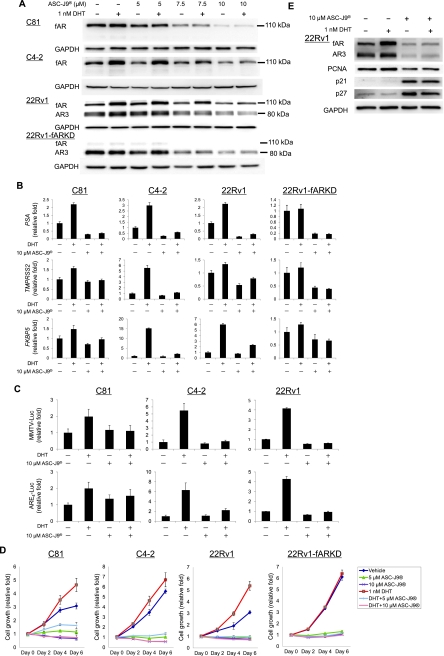
We then determined the effects of ASC-J9 on the expression of fAR and AR3 in C81, C4-2, CWR22Rv1, and CWR22Rv1-fARKD cells. We found that ASC-J9 was able to degrade fAR and AR3 in a dose-dependent manner in various human PCa cells (Figure 3A). The nuclear and cytoplasmic extracts from ASC-J9-treated CWR22Rv1 cells were collected to determine compartmentalized fAR/AR3 expression levels. The results showed that 1 nM DHT could promote fAR nuclear translocation and cotreatment of ASC-J9 could inhibit such nuclear translocation (Figure W1A). AR3 predominantly located in the nuclear compartment and ASC-J9 could also inhibit its amounts (Figure W1A).
Figure 3.
ASC-J9 suppresses AR-targeted genes, AR transactivation, and cell growth through AR degradation in CRPC cells. (A) Degradation of fAR and AR3 by ASC-J9. CRPC cells were treated with 5, 7.5, and 10 µM ASC-J9 in the presence or absence of 1 nM DHT for 24 hours. The expression of fAR and AR3 was evaluated by Western blot analysis in C81 (upper panel), C4-2 (middle panel), CWR22Rv1 (lower panel), and CWR22Rv1-fARKD (bottom panel). (B) Reduction of AR-targeted gene expression after ASC-J9 treatment. C81, C4-2, CWR22Rv1, and CWR22Rv1-fARKD cells were treated with 10 µM ASC-J9. The expressions of AR-targeted genes PSA (upper panel), TMPRSS2 (middle panel), and FKBP5 (lower panel) were determined by real-time PCR analysis. (C) Suppressive effect of ASC-J9 on AR transactivation in CRPC cells. AR transactivational activity was monitored by transfection of MMTV-Luc (upper panel) or ARE4-Luc (lower panel) into C81, C4-2, and CWR22Rv1 cells and treating with 10 µM ASC-J9. The results were normalized to Renilla luciferase and presented as fold difference between groups. (D) Effect of ASC-J9 on cell growth was determined by MTT assay. (E) Elevated cell cycle regulators p21 and p27 in ASC-J9-treated CWR22Rv1 cells leading to inhibition of cell growth. The expressions of fAR/AR3, proliferating cell nuclear antigen (PCNA), p21, p27, and GAPDH were evaluated by Western blot analysis. Data presented are from at least three independent experiments.
In contrast, MDV3100 failed to degrade fAR and AR3 in CWR22Rv1 cells (Figure W2A). Furthermore, we examined the mRNA of fAR and AR3 in CWR22Rv1 cells after ASC-J9 treatment and found that there are few changes compared with vehicle- or DHT-treated group (Figure W2B), suggesting that ASC-J9 mainly targets AR protein degradation.
Next, we examined the effects of ASC-J9 on fAR- and AR3-mediated targeted gene expression. One nanomolar of DHT, the androgen concentration of human prostate tissue after ADT [19], could induce the expression of AR-targeted genes (PSA, TMPRSS2, and FKBP5) in C81, C4-2, and CWR22Rv1 cells but not in CWR22Rv1-fARKD cells (Figure 3B). Addition of ASC-J9 could then suppress 1 nM DHT-induced AR-targeted gene expression in these three cell lines (Figure 3B). Moreover, ASC-J9 could also effectively suppress AR-targeted genes in CWR22Rv1-fARKD cells (Figure 3B), suggesting that ASC-J9 could also degrade AR3 resulting in suppressed AR3-mediated transcription activity. Interestingly, we also found that ASC-J9 was able to inhibit AR3 target genes Akt1 [9] and c-Myc [20] in CWR22Rv1 and CWR22Rv1-fARKD cells (Figure W3A). The qPCR primer sequences of AR canonical-targeted genes (PSA, TMPRSS2, and FKBP5), AR3-targeted genes (Akt1 and c-Myc), AR, and AR3 are provided in Figure W4. Using MMTV and ARE4 luciferase assays, AR transactivational activity was suppressed by ASC-J9 treatment in the presence of 1 nM DHT (Figure 3C).
We also assessed the growth effects of C81, C4-2, and CWR22Rv1 cells with 5 or 10 µM ASC-J9 treatments. The results showed that 1 nM DHT can promote cell growth and ASC-J9 significantly suppressed the DHT-induced cell growth in all three PCa cell lines (Figure 3D). We also observed that ASC-J9 displayed better growth suppression than MDV3100 did in CWR22Rv1 cells (Figure W2C). More importantly, treatment with 5 µM ASC-J9 did not inhibit the cell growth of AR-negative cells (PC-3 and DU-145) (Chang et al., unpublished observations, 2012).
In contrast, although 1 nM DHT failed to induce cell growth in CWR22Rv1-fARKD cells, ASC-J9 could still significantly suppress its growth (Figure 3D, right). The potential mechanisms underlying the antiproliferative effects of ASC-J9 were determined using Western blot analysis to detect proliferation marker (proliferating cell nuclear antigen) and cyclin-dependent kinase inhibitors p21 and p27, suggesting that ASC-J9 may use the up-regulation of p21 and p27 to inhibit the cell growth in CWR22Rv1 cells (Figure 3E). The results were further confirmed using CWR22Rv1-fARKD cells with ASC-J9 treatment (Figure W5A), suggesting that targeting fAR either by shRNA or ASC-J9 could upregulate p27 expression leading to suppressed AR-mediated cell growth, yet degradation of AR3 in CWR22Rv1-fARKD cells did not display the significant changes of p27 expression (Figure W5A).
Together, results from Figures 3 and W3A suggested that, although 1 nM DHT failed to induce AR-mediated targeted genes expression and cell growth in CWR22Rv1-fARKD cells, the existence of AR3 and those AR3-targeted genes could still play important roles to maintain cell growth in CWR22Rv1-fARKD cells. Addition of ASC-J9 to degrade AR3, which resulted in the suppression of those AR3-targeted genes and cell growth in CWR22Rv1-fARKD cells, not only pointed out the important contribution of AR3 in PCa progression but also clearly showed that ASC-J9 treatment could suppress both fAR- and AR3-mediated transcriptional activity and growth in PCa cells.
ASC-J9 Suppresses AR-Targeted Genes and Cell Growth in C81/AR3 and C4-2/AR3 Cells
In addition to studying the impacts of ACS-J9 on the endogenous AR3 in CWR22Rv1 cells, we further validated AC-J9 can degrade AR3 and demonstrated this by ectopically delivering AR3 into C81 and C4-2 cells (Figure 4A). We found that the level of those AR-targeted gene expressions is elevated after adding AR3 and that 1 nM DHT treatment further potentiates the AR-targeted gene expressions (Figure 4B). As expected, ASC-J9 could degrade both endogenous fAR and overexpressed AR3 in both C81/AR3 and C4-2/AR3 cells (Figure 4A). This led to the suppression of 1 nM DHT-induced AR-targeted genes expression (Figure 4B) and cell growth in these AR3-overexpressed cells (Figure 4C).
Figure 4.
ASC-J9 suppresses AR-targeted genes and cell growth by degradation of fAR and ectopic AR3 in C81 and C4-2 cells. (A) Determination of fAR and AR3 expression after ASC-J9 treatment. C81 and C4-2 cells infected with pWPI/AR3 or vector alone were treated with 10 µM ASC-J9 and vehicle or 1 nM DHT for 24 hours. AR expression was evaluated by Western blot analysis. (B) Expressions of AR-targeted genes in AR3 overexpressed cells. C81 and C4-2 cells with ectopically expressed AR3 were treated with ASC-J9. The expressions of AR-targeted genes, such as PSA, TMPRSS2, and FKBP5, were evaluated by real-time PCR. (C) Effects of ASC-J9 on cell growth of C81/Vector, C81/AR3, C4-2/ Vector, and C4-2/AR3 cells were examined by MTT assay. Data presented are from at least three independent experiments.
AR3 Plays a Critical Role to Promote Cell Growth and AR-Targeted Gene Expression in CWR22Rv1/CWR22Rv1-fARKD Cells
To further confirm whether AR3 contributes to cell growth at castration conditions, the lentivirus carrying shAR3 or scrambled control was transduced into CWR22Rv1 and CWR22Rv1-fARKD cells to specifically suppress AR3 expressions (Figure 5A). As shown in Figures 5B and W6A, shAR3 could suppress AR- and AR3-targeted genes, respectively, as well as cell growth in CWR22Rv1 cells (Figure 5C). Importantly, we found that shAR3 could also suppress AR-targeted genes and cell growth in CWR22Rv1-fARKD cells. These results are similar to ASC-J9 treatment in CWR22Rv1-fARKD cells (Figure 3), suggesting that AR3 itself is important to maintain PCa cells growth in such castration conditions.
Figure 5.
Knocking-down AR3 suppresses AR-targeted genes and cell growth in CWR22Rv1 and CWR22Rv1-fARKD cells. (A) Reduction of AR3 expression after delivery of shAR3. We evaluated the shAR3 knockdown efficiency by Western blot analysis in CWR22Rv1 and CWR22Rv1-fARKD cells. (B) Examination of AR-targeted gene expressions after AR3 knockdown. CWR22Rv1 and CWR22Rv1-fARKD cells with scramble control or shAR3 expression were used to detect AR-targeted gene expression by real-time PCR. (C) Effect of AR3 on cell growth of CWR22Rv1 and CWR22Rv1-fARKD cells. CWR22Rv1 and CWR22Rv1-fARKD cells infected with control (solid line) or shAR3 (dashed line) were treated with 1 nM DHT and ASC-J9 followed by MTT cell growth assay. Data presented are from at least three independent experiments.
Collectively, these results of Figures 3 to 5 suggested that AR3 by itself might contribute to the AR-targeted gene expression and AR mediated PCa cell growth, and ASC-J9 could inhibit both fAR- and AR3-mediated gene expression and cell growth in various CRPC cells.
ASC-J9 Suppresses Tumor Growth of CWR22Rv1 Cells in Castrated Mice
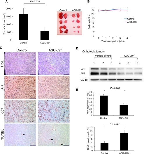
Based on the previously mentioned results, we further evaluated the effects of ASC-J9 in vivo, especially at the castration-resistant stage. We found that even with complete ADT in mice through surgical castration, the orthotopically implanted CWR22Rv1 cells with vehicle treatment were still able to grow into tumors of significant size (Figure 6A). However, mice treated with ASC-J9 had relative smaller tumors compared to those with vehicle injection (Figure 6A). We also found that the body weight was comparable in mice either from vehicle control or ASC-J9-treated (Figure 6B), suggesting little apparent adverse event of ASC-J9.
Figure 6.
Therapeutic effect of ASC-J9 in vivo. (A) Evaluation of tumor volumes of CWR22Rv1 xenografts after ASC-J9 treatment. CWR22Rv1 cells were implanted into anterior prostates of castrated nude mice (four mice each group) and ASC-J9 was intraperitoneally injected for 4 weeks. Orthotopic tumors were harvested (eight tumors each group). Tumor volumes were measured and presented as mean ± SEM. Comparison among groups was performed using Student's t test. (B) Body weight determination during ASC-J9 treatment. Body weights were weekly measured in vehicle- or ASC-J9-injected mice. (C) Histologic examination of tumor tissues after ASC-J9 treatment. The tissues of xenografted tumors were processed by hematoxylin-eosin (HE) staining and immunohistochemical staining for AR, Ki67, and TUNEL. Representative data shown are from four tumors in the ASC-J9 and control groups. Arrows indicate TUNEL-positive cells. Scale bar, 50 µm. (D) Decreased AR expression of CWR22Rv1-xenografted tumors after ASC-J9 treatment. Proteins were harvested from three tumors of each group. AR expression was evaluated by Western blot analysis. (E) Quantification of Ki67 and TUNEL-positive cells in xenografted tumors. The ratio of immunostained positive cells (%) was calculated by the formula: (the number of positive cells/total number of counted cells) x 100. Comparisons among groups were performed using Student's t test.
We performed immunostaining with anti-AR antibody on tumors and found that the AR intensity was reduced in ASC-J9-treated tumors (Figure 6C). Western blot analysis further confirmed that ASC-J9 could degrade both fAR and AR3 in these xenografted tumors in vivo (Figure 6D). In addition, Ki67 staining showed that ASC-J9-treated tumors had significantly decreased Ki67-positive cells (Figure 6, C and E [upper panels]). Finally, TUNEL assay also showed ASC-J9-treated tumors displayed increased apoptotic cells, which may also contribute to tumor growth suppression of these xenografts (Figure 6, C [arrows] and E [lower panel]).
Together, results from Figure 6A to E demonstrated that ASC-J9 treatment might represent the first therapeutic approach that could further suppress CRPC growth in complete androgen-deprived conditions through degradation of fAR and AR3.
Discussion
CWR22 xenografted tumor was established from human primary prostate tumor in the patient with bone metastases [21,22]. CWR22Rv1 is a CRPC cell line derived from the CWR22R subline, which was isolated from the recurrent tumor of the androgen-dependent CWR22 cells in castrated mice [23,24]. The origin of CWR22Rv1 cells was different from other CRPC cell lines, such as C4-2 or C81, that were from metastatic lymph nodes of mice xenografted with human PCa cell line, LNCaP [25–28]. CWR22Rv1 expresses an exon 3-duplicated fAR in addition to the multiple spliced forms [7–9,29]. Recent studies [30] revealed a duplicated AR locus mediated by repetitive elements, which may account for the aberrantly spliced forms of AR. The truncated receptor has been suggested to be derived from two possible pathways, the aberrant splicing as is the case for AR3 and protease cleavage of exon 3-duplicated fAR [29,31]. A different CWR22-derived relapsed cell line, CWR22R1 [32], also displayed abundant truncated AR [9]. Thus, in the evolution of CRPC such as with CWR22, receptor truncation typified by AR3 seems to be a dominant underlying mechanism. Although the detailed truncation points may be different, most the reported species lack the LBD, and are expected not to respond to androgen and antiandrogens. As such, drugs which target the LBD may not work as effectively. The present study is designed to investigate the efficacy of drugs that target degradation of both fAR and the truncated receptor.
In this study, we used various PCa cell lines, including C81, C4-2, and CWR22Rv1 cells, as well as C81/AR3, C4-2/AR3, and CWR22Rv1-fARKD cells to study AR3 in vitro function. The expression level of fAR and AR3 in these PCa cells was different: in C81 and C4-2 cells, most AR expression was fAR (fAR ⋙ AR3). In C81/AR3 and C4-2/AR3 cells, fAR expression level was equivalent to AR3 (fAR = AR3). In contrast, AR3 expression level was higher than fAR (fAR < AR3) in CWR22Rv1 cells, but most AR expression was AR3 in CWR22Rv1-fARKD cells (fAR ⋘ AR3). By characterizing these various PCa cells with differential expression ratios of fAR to AR3, we concluded that AR3 plays a critical role to promote PCa cell growth at some selective stages, which is similar to early findings showing that overexpression of AR3 in LNCaP cells promoted the cell growth [9]. Furthermore, our data showed DHT-induced AR-targeted genes were obviously enhanced with addition of AR3 in C81 and C4-2 cells, suggesting that AR3 might be able to cooperate with fAR to promote DHT-induced AR-targeted genes. This is in agreement with a previous report showing that constitutively active AR splice variants (AR-V7) required fAR to promote AR-targeted genes and cell growth in LNCaP cells in the presence of androgen [12].
However, our data also showed that cell growth of CWR22Rv1-fARKD was substantially increased compared to that of CWR22Rv1 cells under androgen-free conditions. Specifically knocking down AR3 in CWR22Rv1 and CWR22Rv1-fARKD cells resulted in significant growth inhibition, suggesting that AR3 might have dual roles: AR3 by itself might be able to promote PCa cell growth particularly in the absence of androgen at some selective PCa stages and the other role is that AR3 could cooperate with fAR to modulate AR-targeted genes and cell growth in the presence of androgen at many other PCa stages. These conclusions strengthened our central hypothesis and led us to believe that it is necessary to target both fAR and AR3 to have better therapeutic efficacy, especially at castration-resistant stages when AR3 expression is increased.
We demonstrated that Casodex failed to suppress the cell growth of CWR22Rv1 cells, which is in agreement with an early report showing that flutamide, another antiandrogen, had little suppressive effect on the transactivation of ARv567es (another AR splice variant with deletion of AR exon 5–7 regions) [10]. In this present study, we provided a new therapeutic approach through using ASC-J9, which was able to degrade both fAR and AR3 leading to the suppression of their mediated targeted genes and cell growth in various CRPC cells in vitro and in vivo.
In conclusion, our data suggest that AR3 may have its essential roles to promote PCa growth at selective PCa stages and targeting both fAR- and AR3-mediated cell growth through ASC-J9 may become a new useful therapeutic strategy to target PCa cells, which express AR splice variants such as AR3.
Supplementary Methods
Cell and Reagents
Human PCa cell lines CWR22Rv1 and CWR22Rv1-fARKD were used. The antibodies for AR (N-20), PARP-1, α-tubulin, p27, and GAPDH were purchased from Santa Cruz Biotechnology. MDV3100 was purchased from SelleckChem (Houston, TX) and ASC-J9 (5-hydroxy-1,7-bis(3,4-dimethoxyphenyl)-1,4,6-heptatrien-3-one) is a gift from AndroScience. The NE-PER nuclear and cytoplasmic extraction reagents purchased from Thermo Scientific (Rockford, IL) were used to separate nuclear and cytoplasmic fractions.
Cell Growth Assay
Cells were treated with vehicle, 1 nM DHT, 10 µM ASC-J9, and 5 or 10 µM MDV3100 in 10% CD-FBS medium. The media were replenished every other day, and we followed the standard MTT assay protocols.
Western Blot Analysis and Quantitative Real-time PCR
Cells were cultured and treated with or without 10 µM MDV3100 or 10 µM ASC-J9 for 24 hours in 10% CD-FBS medium. Cell lysates were harvested and subjected to Western blot analysis. Quantitative real-time PCR (qPCR) was performed in triplicate with a Bio-Rad iCycler system, and mRNA levels of fAR, AR3, Akt1, c-Myc, and GAPDH were measured.
Acknowledgments
The authors thank Karen Wolf for assistance in article preparation and Yun Qiu (University of Maryland) and Jun Luo (Johns Hopkins University) for their gifts of AR3 plasmid and AR-V7 antibody, respectively. The authors also thank Koji Mitsuzuka and Mitsuharu Sasaki (Tohoku University Graduate School of Medicine) for helping collect human PCa samples.
Abbreviations
- AR
androgen receptor
- CRPC
castration-resistant prostate cancer
- ADT
androgen deprivation therapy
- fAR
full-length AR
- shAR3
short-hairpin AR3
Footnotes
ASC-J9 was patented by the University of Rochester, the University of North Carolina, and AndroScience Corp and then licensed to AndroScience Corp. Both the University of Rochester and Chawnshang Chang own royalties and equity in AndroScience Corp. This study was supported by the National Institutes of Health (CA122840 and CA127300), George Whipple Professorship Endowment, and National Science Council, Taiwan Department of Health Clinical Trial, and Research Center of Excellence grant DOH99-TD-B-111-004 (China Medical University, Taichung, Taiwan).
This article refers to supplementary materials, which are designated by Table W1 and Figures W1 to W6 and are available online at www.neoplasia.com.
References
- 1.Jemal A, Siegel R, Xu J, Ward E. Cancer statistics, 2010. CA Cancer J Clin. 2010;60:277–300. doi: 10.3322/caac.20073. [DOI] [PubMed] [Google Scholar]
- 2.Huggins C, Hodges CV. Studies on prostatic cancer: I. The effect of castration, of estrogen and of androgen injection on serum phosphatases in metastatic carcinoma of the prostate. Cancer Res. 1941;1:293–297. doi: 10.3322/canjclin.22.4.232. [DOI] [PubMed] [Google Scholar]
- 3.Eisenberger MA, Blumenstein BA, Crawford ED, Miller G, McLeod DG, Loehrer PJ, Wilding G, Sears K, Culkin DJ, Thompson IM, Jr, et al. Bilateral orchiectomy with or without flutamide for metastatic prostate cancer. N Engl J Med. 1998;339:1036–1042. doi: 10.1056/NEJM199810083391504. [DOI] [PubMed] [Google Scholar]
- 4.Heidenreich A, Aus G, Bolla M, Joniau S, Matveev VB, Schmid HP, Zattoni F. EAU guidelines on prostate cancer. Eur Urol. 2008;53:68–80. doi: 10.1016/j.eururo.2007.09.002. [DOI] [PubMed] [Google Scholar]
- 5.Chang CS, Kokontis J, Liao ST. Molecular cloning of human and rat complementary DNA encoding androgen receptors. Science. 1988;240:324–326. doi: 10.1126/science.3353726. [DOI] [PubMed] [Google Scholar]
- 6.Chen CD, Welsbie DS, Tran C, Baek SH, Chen R, Vessella R, Rosenfeld MG, Sawyers CL. Molecular determinants of resistance to antiandrogen therapy. Nat Med. 2004;10:33–39. doi: 10.1038/nm972. [DOI] [PubMed] [Google Scholar]
- 7.Dehm SM, Schmidt LJ, Heemers HV, Vessella RL, Tindall DJ. Splicing of a novel androgen receptor exon generates a constitutively active androgen receptor that mediates prostate cancer therapy resistance. Cancer Res. 2008;68:5469–5477. doi: 10.1158/0008-5472.CAN-08-0594. [DOI] [PMC free article] [PubMed] [Google Scholar]
- 8.Hu R, Dunn TA, Wei S, Isharwal S, Veltri RW, Humphreys E, Han M, Partin AW, Vessella RL, Isaacs WB, et al. Ligand-independent androgen receptor variants derived from splicing of cryptic exons signify hormone-refractory prostate cancer. Cancer Res. 2009;69:16–22. doi: 10.1158/0008-5472.CAN-08-2764. [DOI] [PMC free article] [PubMed] [Google Scholar]
- 9.Guo Z, Yang X, Sun F, Jiang R, Linn DE, Chen H, Kong X, Melamed J, Tepper CG, Kung HJ, et al. A novel androgen receptor splice variant is up-regulated during prostate cancer progression and promotes androgen depletion-resistant growth. Cancer Res. 2009;69:2305–2313. doi: 10.1158/0008-5472.CAN-08-3795. [DOI] [PMC free article] [PubMed] [Google Scholar]
- 10.Sun S, Sprenger CC, Vessella RL, Haugk K, Soriano K, Mostaghel EA, Page ST, Coleman IM, Nguyen HM, Sun H, et al. Castration resistance in human prostate cancer is conferred by a frequently occurring androgen receptor splice variant. J Clin Invest. 2010;120:2715–2730. doi: 10.1172/JCI41824. [DOI] [PMC free article] [PubMed] [Google Scholar]
- 11.Heinlein CA, Chang C. Androgen receptor in prostate cancer. Endocr Rev. 2004;25:276–308. doi: 10.1210/er.2002-0032. [DOI] [PubMed] [Google Scholar]
- 12.Watson PA, Chen YF, Balbas MD, Wongvipat J, Socci ND, Viale A, Kim K, Sawyers CL. Constitutively active androgen receptor splice variants expressed in castration-resistant prostate cancer require full-length androgen receptor. Proc Natl Acad Sci USA. 2010;107:16759–16765. doi: 10.1073/pnas.1012443107. [DOI] [PMC free article] [PubMed] [Google Scholar]
- 13.Niu Y, Altuwaijri S, Lai KP, Wu CT, Ricke WA, Messing EM, Yao J, Yeh S, Chang C. Androgen receptor is a tumor suppressor and proliferator in prostate cancer. Proc Natl Acad Sci USA. 2008;105:12182–12187. doi: 10.1073/pnas.0804700105. [DOI] [PMC free article] [PubMed] [Google Scholar]
- 14.Mir C, Shariat SF, Van Der Kwast TH, Ashfaq R, Lotan Y, Evans A, Skeldon S, Hanna S, Vajpeyi R, Kuk C, et al. Loss of androgen receptor expression is not associated with pathological stage, grade, gender or outcome in bladder cancer: a large multi-institutional study. BJU Int. 2010;108:24–30. doi: 10.1111/j.1464-410X.2010.09834.x. [DOI] [PubMed] [Google Scholar]
- 15.Miyamoto H, Yang Z, Chen YT, Ishiguro H, Uemura H, Kubota Y, Nagashima Y, Chang YJ, Hu YC, Tsai MY, et al. Promotion of bladder cancer development and progression by androgen receptor signals. J Natl Cancer Inst. 2007;99:558–568. doi: 10.1093/jnci/djk113. [DOI] [PubMed] [Google Scholar]
- 16.Yang Z, Chang YJ, Yu IC, Yeh S, Wu CC, Miyamoto H, Merry DE, Sobue G, Chen LM, Chang SS, et al. ASC-J9 ameliorates spinal and bulbar muscular atrophy phenotype via degradation of androgen receptor. Nat Med. 2007;13:348–353. doi: 10.1038/nm1547. [DOI] [PubMed] [Google Scholar]
- 17.Wu MH, Ma WL, Hsu CL, Chen YL, Ou JH, Ryan CK, Hung YC, Yeh S, Chang C. Androgen receptor promotes hepatitis B virus-induced hepatocarcinogenesis through modulation of hepatitis B virus RNA transcription. Sci Transl Med. 2010;2:32ra35. doi: 10.1126/scitranslmed.3001143. [DOI] [PMC free article] [PubMed] [Google Scholar]
- 18.Ohtsu H, Xiao Z, Ishida J, Nagai M, Wang HK, Itokawa H, Su CY, Shih C, Chiang T, Chang E, et al. Antitumor agents. 217. Curcumin analogues as novel androgen receptor antagonists with potential as anti-prostate cancer agents. J Med Chem. 2002;45:5037–5042. doi: 10.1021/jm020200g. [DOI] [PubMed] [Google Scholar]
- 19.Marks LS, Mostaghel EA, Nelson PS. Prostate tissue androgens: history and current clinical relevance. Urology. 2008;72:247–254. doi: 10.1016/j.urology.2008.03.033. [DOI] [PMC free article] [PubMed] [Google Scholar]
- 20.Zhang X, Morrissey C, Sun S, Ketchandji M, Nelson PS, True LD, Vakar-Lopez F, Vessella RL, Plymate SR. Androgen receptor variants occur frequently in castration resistant prostate cancer metastases. PLoS One. 2011;6:e27970. doi: 10.1371/journal.pone.0027970. [DOI] [PMC free article] [PubMed] [Google Scholar]
- 21.Pretlow TG, Wolman SR, Micale MA, Pelley RJ, Kursh ED, Resnick MI, Bodner DR, Jacobberger JW, Delmoro CM, Giaconia JM, et al. Xenografts of primary human prostatic carcinoma. J Natl Cancer Inst. 1993;85:394–398. doi: 10.1093/jnci/85.5.394. [DOI] [PubMed] [Google Scholar]
- 22.Wainstein MA, He F, Robinson D, Kung HJ, Schwartz S, Giaconia JM, Edgehouse NL, Pretlow TP, Bodner DR, Kursh ED, et al. CWR22: androgen-dependent xenograft model derived from a primary human prostatic carcinoma. Cancer Res. 1994;54:6049–6052. [PubMed] [Google Scholar]
- 23.Nagabhushan M, Miller CM, Pretlow TP, Giaconia JM, Edgehouse NL, Schwartz S, Kung HJ, de Vere White RW, Gumerlock PH, Resnick MI, et al. CWR22: the first human prostate cancer xenograft with strongly androgen-dependent and relapsed strains both in vivo and in soft agar. Cancer Res. 1996;56:3042–3046. [PubMed] [Google Scholar]
- 24.Sramkoski RM, Pretlow TG, II, Giaconia JM, Pretlow TP, Schwartz S, Sy MS, Marengo SR, Rhim JS, Zhang D, Jacobberger JW. A new human prostate carcinoma cell line, 22Rv1. In Vitro Cell Dev Biol Anim. 1999;35:403–409. doi: 10.1007/s11626-999-0115-4. [DOI] [PubMed] [Google Scholar]
- 25.Horoszewicz JS, Leong SS, Chu TM, Wajsman ZL, Friedman M, Papsidero L, Kim U, Chai LS, Kakati S, Arya SK, et al. The LNCaP cell line—a new model for studies on human prostatic carcinoma. Prog Clin Biol Res. 1980;37:115–132. [PubMed] [Google Scholar]
- 26.van Bokhoven A, Varella-Garcia M, Korch C, Johannes WU, Smith EE, Miller HL, Nordeen SK, Miller GJ, Lucia MS. Molecular characterization of human prostate carcinoma cell lines. Prostate. 2003;57:205–225. doi: 10.1002/pros.10290. [DOI] [PubMed] [Google Scholar]
- 27.Wu HC, Hsieh JT, Gleave ME, Brown NM, Pathak S, Chung LW. Derivation of androgen-independent human LNCaP prostatic cancer cell sublines: role of bone stromal cells. Int J Cancer. 1994;57:406–412. doi: 10.1002/ijc.2910570319. [DOI] [PubMed] [Google Scholar]
- 28.Lin MF, Meng TC, Rao PS, Chang C, Schonthal AH, Lin FF. Expression of human prostatic acid phosphatase correlates with androgen-stimulated cell proliferation in prostate cancer cell lines. J Biol Chem. 1998;273:5939–5947. doi: 10.1074/jbc.273.10.5939. [DOI] [PubMed] [Google Scholar]
- 29.Tepper CG, Boucher DL, Ryan PE, Ma AH, Xia L, Lee LF, Pretlow TG, Kung HJ. Characterization of a novel androgen receptor mutation in a relapsed CWR22 prostate cancer xenograft and cell line. Cancer Res. 2002;62:6606–6614. [PubMed] [Google Scholar]
- 30.Li Y, Alsagabi M, Fan D, Bova GS, Tewfik AH, Dehm SM. Intragenic rearrangement and altered RNA splicing of the androgen receptor in a cell-based model of prostate cancer progression. Cancer Res. 2011;71:2108–2117. doi: 10.1158/0008-5472.CAN-10-1998. [DOI] [PMC free article] [PubMed] [Google Scholar]
- 31.Chen H, Libertini SJ, Wang Y, Kung HJ, Ghosh P, Mudryj M. ERK regulates calpain 2-induced androgen receptor proteolysis in CWR22 relapsed prostate tumor cell lines. J Biol Chem. 2010;285:2368–2374. doi: 10.1074/jbc.M109.049379. [DOI] [PMC free article] [PubMed] [Google Scholar]
- 32.Gregory CW, Johnson RT, Jr, Mohler JL, French FS, Wilson EM. Androgen receptor stabilization in recurrent prostate cancer is associated with hypersensitivity to low androgen. Cancer Res. 2001;61:2892–2898. [PubMed] [Google Scholar]
Associated Data
This section collects any data citations, data availability statements, or supplementary materials included in this article.


