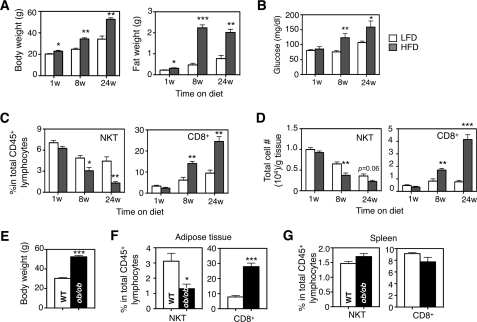
FIGURE 1.
Abundance of adipose NKT cells decreases with HFD feeding in two obese mouse models. Body, epididymal fat weights (A), and fasting glucose levels (B) of wild type C57BL/6 male mice under HFD (60%) for 1–24 weeks (w) were compared with age-matched male mice on LFD (13% fat). n = 12–15 mice each. C, percentages of NKT and CD8+ T lymphocytes in total lymphocytes in adipose tissue during HFD feeding compared with age-matched LFD cohort. D, total cell numbers of various T lymphocytes per g of adipose tissue during HFD feeding. C and D, n = 12 mice with three repeats. E, body weight of adult 36–40-week-old ob/ob mice. F and G, percentages of NKT and CD8+ T cells in adipose tissue (F) and spleen (G) of ob/ob mice compared with age-matched WT lean animals. n = 10 mice in each cohort, two repeats. Values represent mean ± S.E. *, p < 0.05; **, p < 0.01; and ***, p < 0.005.

