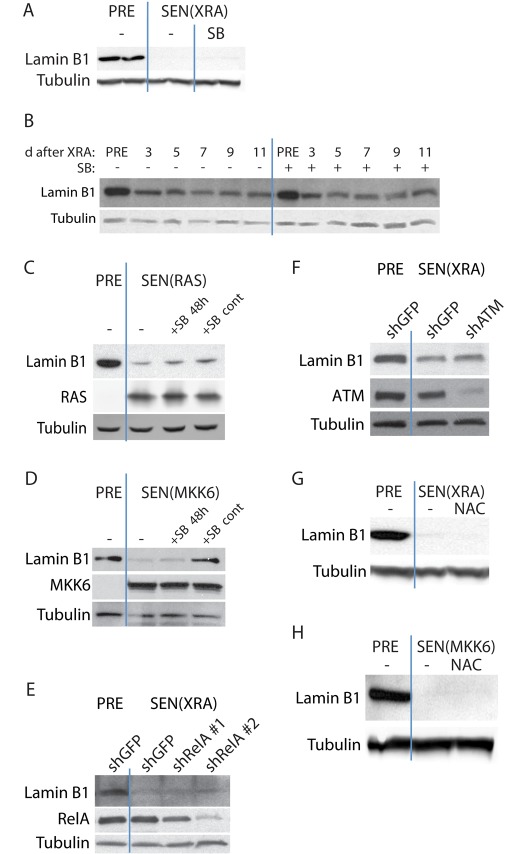
FIGURE 2:
Lamin B1 loss at senescence is independent of p38 MAPK, NF-κB, ATM, and ROS signaling. (A) p38 MAPK inhibition does not reverse lamin B1 decline in DNA damage–induced senescence. The p38 MAPK inhibitor SB203580 (SB; 10 μM) was added to SEN(XRA) HCA2 cells for 48 h. Whole-cell lysates were analyzed by Western blotting. PRE cells were mock irradiated. (B) p38 MAPK inhibition does not prevent lamin B1 decline in DNA damage–induced senescence. SB was added to HCA2 cells before XRA. Cells were mock irradiated (PRE) or irradiated (XRA). Whole-cell lysates collected at the indicated time points were analyzed by Western blotting. SB was replaced daily. (C) p38 MAPK inhibition does not reverse or prevent RAS-induced lamin B1 decline. HCA2 cells were infected with a lentivirus expressing oncogenic RASV12 and allowed to senesce (SEN(RAS)). SB was not added (–), added 8 d after infection for 48 h (+SB 48 h), or added before infection and maintained for 10 d (+SB cont). Presenescent controls (PRE) were infected with an insertless vector. Whole-cell lysates were analyzed by Western blotting. (D) p38 MAPK inhibition prevents but does not reverse MKK6-induced lamin B1 decline. HCA2 cells were infected with a lentivirus expressing MKK6EE and allowed to senesce (SEN(MKK6)). SB was not added (–), added 8 d after infection for 48 h (+SB 48 h), or added before infection and maintained for 10 d (+SB cont). Presenescent controls (PRE) were infected with an insertless vector. Whole-cell lysates were analyzed by Western blotting. (E) RelA depletion does not prevent DNA damage–induced lamin B1 decline. HCA2 cells were infected with a lentivirus expressing either of two shRNAs against RelA (shRelA) or GFP (shGFP; control) and selected. Cells were then irradiated and allowed to senesce (SEN(XRA)). Presenescent controls (PRE) were mock irradiated. Whole-cell lysates were analyzed by Western blotting. (F) ATM depletion does not prevent DNA damage–induced lamin B1 decline. HCA2 cells were infected with a lentivirus expressing an shRNA against ATM (shATM) or GFP (shGFP; control) and selected. Cells were then irradiated and allowed to senesce (SEN(XRA)). Presenescent controls (PRE) were mock irradiated. Whole-cell lysates were analyzed by Western blotting. (G) ROS inhibition does not prevent DNA damage–induced lamin B1 decline. NAC (10 mM) was added to HCA2 cells before irradiation and maintained until whole-cell lysates were collected 10 d after XRA (SEN(XRA)). Presenescent controls (PRE) were mock irradiated. NAC was replaced daily. Whole-cell lysates were analyzed by Western blotting. (H) ROS inhibition does not prevent MKK6-induced lamin B1 decline. NAC (10 mM) was added to HCA2 cells before infection and continued until sample collection. Whole-cell lysates were collected 10 d after infection with a lentivirus expressing MKK6EE (SEN(MKK6)). Presenescent controls (PRE) were infected with a lentivirus lacking an insert. NAC was replaced daily. Whole-cell lysates were analyzed by Western blotting.

