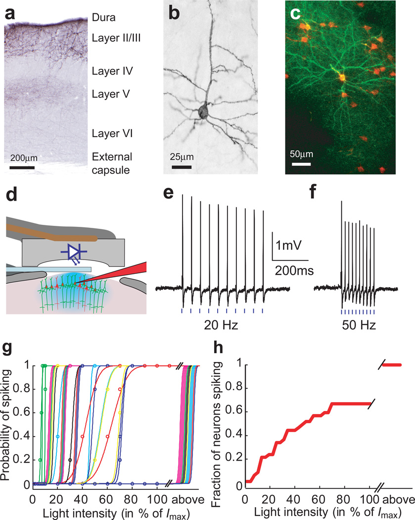
Figure 1. ChR2-assisted photostimulation of layer 2/3 barrel cortex neurons in vivo.
(a) Coronal section through the electroporated mouse somatosensory cortex after immunohistochemical staining for ChR2-GFP. (b) Individual layer 2/3 neuron, side view. (c) Maximum value projection (top view) of an image stack in vivo (see Supplementary Movie 1) showing layer 2/3 neurons expressing ChR2-GFP and cytosolic RFP. (d) Schematic of the recording geometry.. (e) Action potentials recorded from one ChR2-GFP-positive neuron. Blue bars indicate photostimuli (1 ms duration, 11.6 mW/mm2, 20 Hz). (f) Same as e, 50 Hz. (g) Probability of spiking as a function of light intensity (1 ms duration, 5 repetitions per condition, 15 sec between stimuli) (Imax = 11.6 mW/mm2). Each line corresponds to a different neuron, each color to a different animal. Neurons that could only be driven with photostimuli longer than 1ms were pooled at the far right (above). (h) Cumulative fraction of recorded neurons firing at various threshold intensity levels (computed from the data in g).

