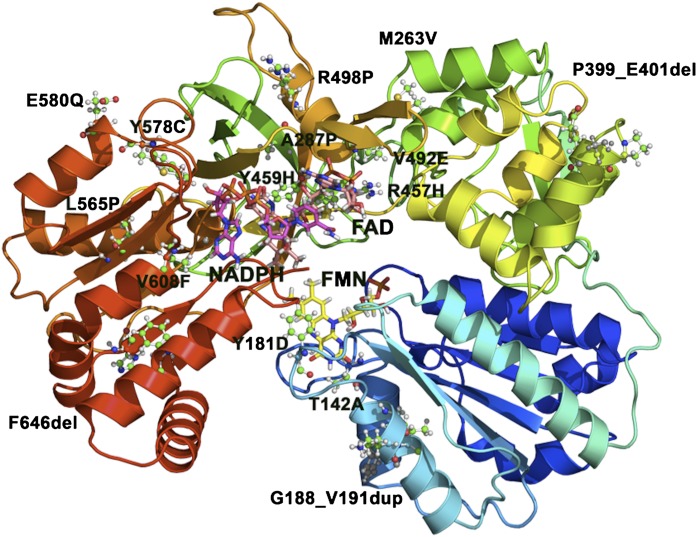
Fig. 10.
Mutations in POR. The structure of human POR, with missense mutations identified in patients with POR deficiency, is shown as a ribbon model. Mutations are shown as ball-and-stick models, whereas FMN (yellow), FAD (orange), and NADPH (magenta) are shown as stick models. Mutations that result in a stop codon and/or cause truncations in POR are not shown.

