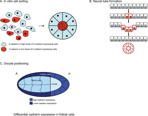
Figure 4. The role of cadherin in cell sorting and positioning.
(A) Differential type or levels of cadherin expression on cells drive cell sorting in vitro in cell (re)aggregation assays. (B) During the formation of the neural tube E-cadherin is switched off in a subset of ectodermal cells, whereas N-cadherin expression is turned on in these cells (red cell membranes) driving segregation of these cell populations. Other in vivo examples are neural crest cell migration and positioning and segregation of motor neuron cells. (C) Drosophila oocyte positioning where differential cadherin expression in the follicle cells is crucial to properly position the oocyte at the posterior end of the embryo. For detailed description see text.

