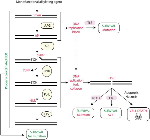
Figure 5. Cellular processing and repair of 3meA lesions in DNA.
Monofunctional methylating agents induce the formation of 3-methyladenine (3meA) lesions, which are excised by the alkyladenine DNA glycosylase (AAG) to generate an apurinic/apyridinic site (AP). The apurinic/apyridinic endonuclease (APE) incises the DNA backbone immediately 5′ to the AP site generating a single strand break (SSB) containing a 3-hydroxyl (3′OH) and a 5′-deoxyribose phosphate (5′dRP). DNA polymerase β (Pol β) removes the 5′dRP moiety through intrinsic lyase activity and fills in the resulting gap. The nick is then sealed by DNA ligase (LIG) to complete base excision repair (BER). If BER is inhibited or downstream steps of BER are limiting, then toxic intermediates can accumulate leading to DNA replication blocks, replication fork collapse, double strand breaks (DSBs) and eventually cell death. The translesion DNA synthesis (TLS), homologous recombination (HR) and non-homologous end-joining (NHEJ) pathways can bypass or repair secondary damage caused by alkyl lesions to promote cell survival at the expense of mutation or sister chromatid exchange (SCE).

