Figure 3.
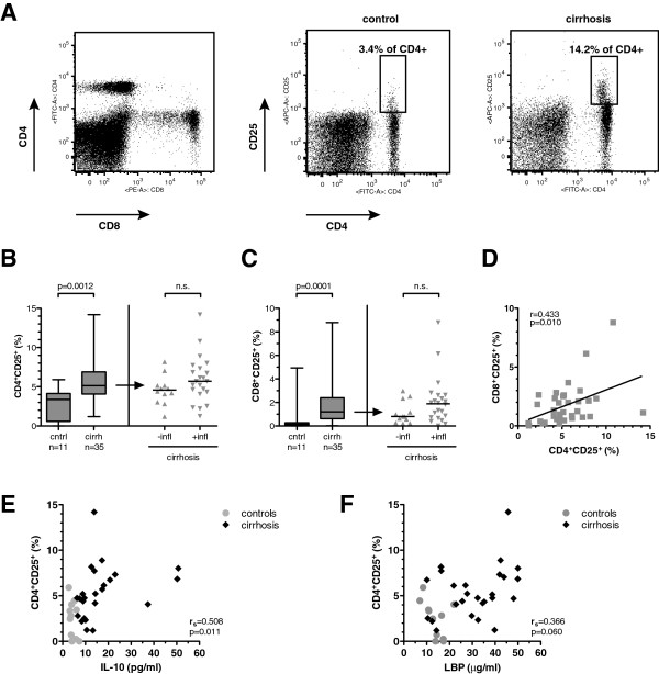
Increased fraction of CD25+ T cells in cirrhosis. (A) Gating strategy (left) and representative plots of CD25 expression on CD4 T cells in a healthy control (middle) and a patients with cirrhosis (right). Panels B and C display distribution and median of CD25-positive CD4+ T cells (B) and CD8+ T cells (C) in healthy controls and patients with cirrhosis (left) and in cirrhotics stratified for inflammation (right). P values in Mann–Whitney U test are indicated; n.s. = not significant. (D) Correlation of CD4+ CD25+ T cells and CD8+ CD25+ T cells in cirrhotic individuals. Linear regression curve, Pearson product–moment correlation coefficient R and P value are indicated. Panels E and F show the correlation of percentage of CD25-positive CD4+ T cells with IL-10 serum concentration (E) and LBP serum concentration (F) in patients with cirrhosis (black diamonds) and controls (grey circles), respectively. Spearman’s correlation coefficient Rs and P value are indicated for the cirrhotic patients.

