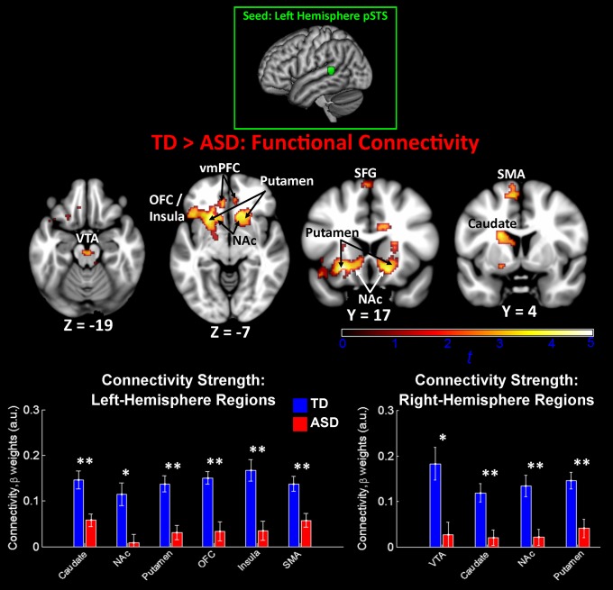
Fig. 2.
Between-group functional connectivity results for left-hemisphere voice-selective cortex. Group differences for the TD>ASD contrast indicated ASD underconnectivity between left-hemisphere pSTS and structures of the reward network, including the VTA, nucleus accumbens (NAc), insula, and OFC. No voxels showed significant connectivity for the ASD>TD contrast. The seed used in this analysis was a 6-mm sphere centered in left-hemisphere pSTS at MNI coordinates [−63, −42, 9] (23). Images are thresholded at P < 0.01 for voxel height and an extent of 100 voxels. Mean connectivity differences between TD children and children with ASD are plotted in the bar graphs for six left-hemisphere and four right-hemisphere regions (error bars represent SEM). SFG, superior frontal gyrus; SMA, supplementary motor area.

