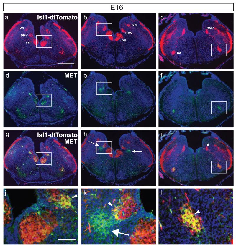
Fig. 2.
MET protein expression in neurons residing in DMV, nXII, and nTS. Micrographs show MET immunofluorescence in a series of coronal sections harvested from Isl1cre/Rosa-tdTomatofx/+ mice at E16. a-c, endogenous tdTomato fluorescence, expressed under the control of Isl1cre at 3 successive levels of the medulla; d-f, MET immunostaining on the same sections; g-i overlay of tdTomato and MET immunofluorescence. Regions in white boxes are presented at higher magnification in j-l. VN, vestibular nucleus. Arrowheads indicate subsets of MET+/tdTomato+ neurons in nX (j and k) and in nA (l). Arrows point to a subset of MET+/tdTomato− neurons located lateral to DMV. An almost complete overlap of tdTomato and MET immunofluorescence is observed in the nA (l). No MET-immunoreactivity was found in the main VN (g) and in rostral part of DMV (i), which is indicated by an asterisk. Scale bar, a-i: 50 μm, j-l: 10 μm.

