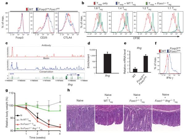
Figure 4. Restoration of Foxo1-deficient Treg cell function in the absence of IFN-γ.
a, Flow cytometric analysis of Foxp3, CD25 and CTLA4 expression in lymph node Treg cells from 20-day-old wild-type and Foxp3creFoxo1fl/fl mice. b, Suppression of wild-type naive CD4+ T cells, labelled with the cytosolic dye carboxyfluorescein diacetate succinimidyl ester (CFSE) (responding T cells (Tresp)), by wild-type and Foxo1-deficient (Foxo1−/−) Treg cells. Tresp cell division was assessed by CFSE dilution at the indicated ratios of cell numbers between Treg cell and Tresp cells. c, Foxo1-bound regions in the Ifng locus. d, Foxo1 binding to the ChIP-seq peak was confirmed by ChIP-quantitative (q)PCR, and is presented relative to enrichment by immunoprecipitation with isotype-matched control antibody. e, Expression of IFN-γ mRNA in Treg cells from wild-type and Foxp3creFoxo1fl/fl mice was determined by qPCR. f, Peripheral lymph node T cells from 20-day-old wild-type and Foxp3creFoxo1fl/fl mice were stimulated with PMA and ionomycin for 4 h. The expression of IFN-γ in Treg cells was determined by intracellular staining. g, h, Naive T cells (N) were transferred to Rag1−/− mice alone or in combination with wild-type, Foxo1−/− or Foxo1−/− Ifng−/− Treg cells. Changes in body weight of Rag1−/− recipients over time are shown (n=4) (g). Haematoxylin and eosin staining of colon sections from Rag1−/− mice at 4 weeks after the transfer (h). Error bars represent s.d.; results are representative of at least three independent experiments.

