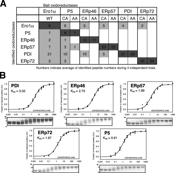
Figure 6.
PDI works as a penultimate electron acceptor in the Ero1-α–mediated oxidation cascade. (A) Oxidoreductase mutants (CA or AA) and Ero1-α(WT) with FLAG tag were expressed in HEK293T cells, and anti-FLAG immunoprecipitates were analyzed by direct nanoflow liquid chromatography coupled with tandem mass spectrometry. Reproducibly identified oxidoreductases from four independent trials are listed. Each number indicates the identified peptide number of each protein in an individual experiment. Light and dark shading indicate identified prey and bait peptides, respectively. (B) Free sulfhydryl groups of the cysteine residues were modified with mPEG2000-mal after incubation with different [GSH]2/[GSSG] ratios in a buffer containing 0.1 mM GSSG and varying concentrations of GSH (0.05–10 mM) under a nonoxidative atmosphere at 25°C followed by SDS-PAGE and CBB staining. The apparent equilibrium constants between oxidoreductases and glutathione were determined by the nonlinear least square fitting of the data (Fig. S4, A–D). Keq values were determined from at least three independent trials as follows: 3.02 ± 0.14 (PDI, correlation coefficient: 0.985), 2.73 ± 0.10 (ERp46, 0.994), 1.69 ± 0.12 (ERp57, 0.991), 1.67 ± 0.08 (ERp72, 0.994), and 0.91 ± 0.04 (P5, 0.995). A.U., arbitrary unit.

