Fig. 5.
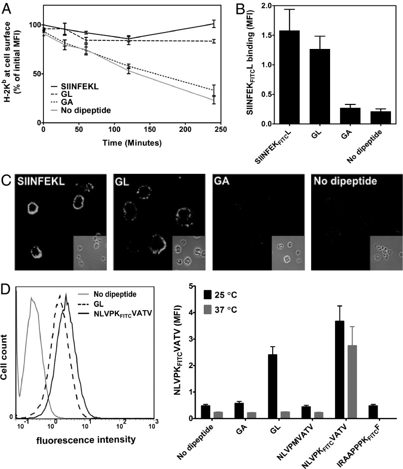
GL stabilizes peptide-receptive class I molecules at the cell surface. (A) BFA decay experiment. RMA-S cells were incubated overnight at 25 °C, then 20 mM GL or GA (or 20 µM SIINFEKL as positive control) were added to the medium, cells were transferred to 37 °C in the presence of BFA, and Kb surface levels were determined at each time point with MAb Y3 and flow cytometry. Averages ± SD (n = 3) are normalized to initial mean fluorescence intensity (MFI). (B) Peptide binding of GL-stabilized cell surface Kb molecules. In an experiment as in A, cells were taken at the 4-h time point, dipeptides were washed off, 1 µM fluorescently labeled SIINFEKFITCL was added, and binding was assessed by flow cytometry. SIINFEKFITCL present during the 4-h incubation was used as positive control (leftmost bar). (C) In an experiment as in A, cell surface levels of Kb after 4 h of incubation at 37 °C were detected with MAb Y3 and immunofluorescence microscopy. Microscope settings are the same for all panels. (D) Cell surface accumulation of A2 molecules. T2 cells were incubated overnight at 25 °C with 20 mM GL, 5 µM NLVPKFITCVATV, or without peptide. After washing with PBS solution, 5 µM NLVPKFITCVATV was added, and binding was detected by flow cytometry (Left). (Right) Comparison of cell surface A2 molecules accumulated overnight at 25 °C (n = 5) and 37 °C (n = 2). Unlabeled NLVPMVATV (20 µM) and the nonspecific peptide IRAAPPPKFITCF were used as negative controls.

