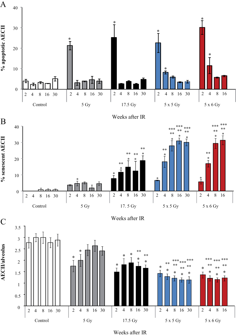
Figure 4.
Extent of senescence and apoptosis in irradiated mouse lungs. C57Bl/6NCr mice were exposed to thoracic irradiation at doses of 0 Gy (control), 5 Gy, 17.5 Gy, 5×6 Gy, or 5×5 Gy. Samples of lung tissue were collected at intervals after irradiation (IR) and in controls (n = 3 per dose and time-point). A) The percentage of type II airway epithelial cell (AECII) cells that stain for apoptosis (TUNEL) was scored by dose and time point in lungs of mice treated with 0 Gy (control), 5 Gy, 17.5 Gy, 5×5 Gy, or 5×6 Gy. B) The percentage of AECII cells that stain for senescence was scored by dose and time point in lungs of mice treated with no IR (control), 5 Gy, 17.5 Gy, 5×6 Gy, and 5×5 Gy. C) The number of AECII cells per alveoli was scored by dose and time point in lungs of mice treated with no IR (control), 5 Gy, or 17.5 Gy, 5×5 Gy, and 5×6 Gy. Bars represent the mean. Error bars represent the standard error. *P < .05 for the comparison of each treatment to controls; **P < .05 for the comparison of irradiated groups to 5 Gy within the same time-point; ***P < .05 by analysis of variance for the comparison of irradiated groups to 17.5 Gy within the same time point. All statistical tests were two-sided.

