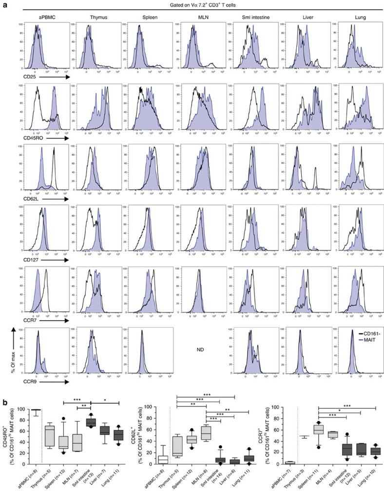
Figure 3. Detailed phenotypic analysis of fetal MAIT cells.
(a) CD25, CD45RO, CD62L, CD127, CCR7 and CCR9 expression was determined on Vα7.2+ CD161hi MAIT cells and Vα7.2+ CD161− T cells from fetal tissues and control adult PBMCs from at least three independent donors. Representative FACS plots are shown. (b) Expression of CD45RO, CD62L and CCR7 on MAIT cells is shown in left, middle and right subpanel, respectively. CD45RO expression was determined in the thymus (n=5), spleen (n=13), mesenteric lymph nodes (MLN) (n=7), small (sml) intestine (n=13), liver (n=7) and lung (n=11). CD62L was assessed in the thymus (n=5), spleen (n=12), MLN (n=8), sml intestine (n=14), liver (n=6) and lung (n=11). CCR7 was determined in the thymus (n=3), spleen (n=11), MLN (n=4), sml intestine (n=12), liver (n=5) and lung (n=10). Box and whisker plot shows median, IQR and the 10th to the 90th percentile. *P<0.05, **P<0.01, ***P<0.001 (Kruskal–Wallis ANOVA followed by Dunn’s post-hoc test).

