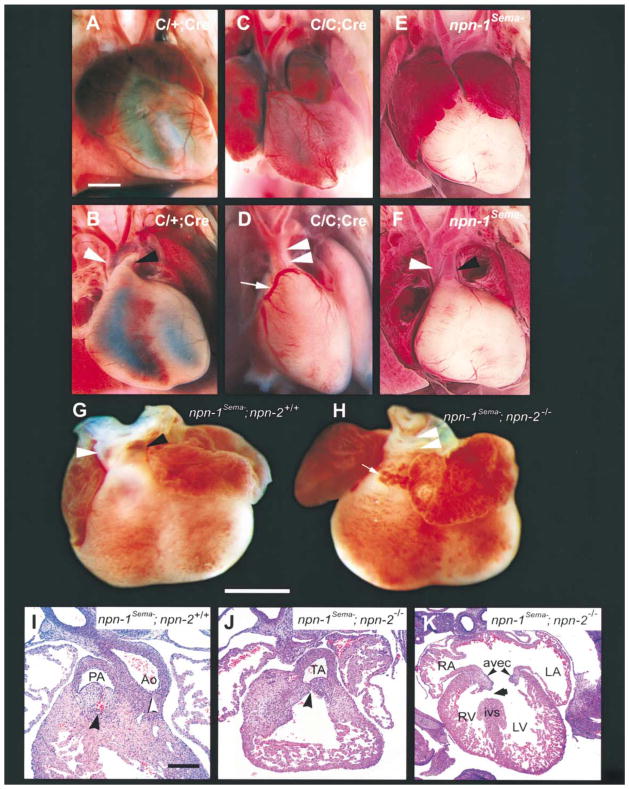
Figure 7. Cardiac Defects in Endothelial Cell-Specific npn-1 Null Mice, npn-1Sema− Mice, and npn-1Sema−;npn-2−/− Double Mutant Mice.
(A–F) Gross examination of the hearts of wild-type (A and B), endothelial cell-specific npn-1 null mice (C/C;Cre) (C and D), and npn-1Sema− mice (E and F). (A and B) Ventral view of a wild-type heart in which normal atria, ventricles, and vessels of the aortic arch can be seen (A). After removal of the anterior half of the atria (B), the right ventricular outflow tract can be clearly seen giving rise to the pulmonary trunk (black arrowhead). The aorta (white arrowhead) is distinctly visible arising posterior to the pulmonary trunk (B). The left anterior descending coronary artery (faint) courses normally through the interventricular sulcus. (C and D) Ventral view of the heart from a C/C;Cre mutant. There is marked right atrial enlargement (C). The ventricular outflow tract shows a common trunk (double arrowhead in [D]) giving rise to both the aorta and the pulmonary trunk (truncus arteriosus). Upon removal of the anterior half of the atria (D) an anomalous origin of the left coronary artery from the right side of the truncus arteriosus can be seen (white arrow), and this misplaced coronary artery crosses over the outflow tract to become the left anterior descending artery. (E and F) Ventral view of the heart of a npn-1Sema− mouse. There is bilateral atrial enlargement (E), but, upon removal of the atria, it is clear that the aorta, pulmonary arteries, and coronary arteries arise normally.
(G–K) Persistent truncus arteriosus in E14.5 npn-1Sema−;npn-2−/− double mutant mice. (G and H) Gross examination of the heart. (G) Ventral view of a heart from an E14.5 npn-1Sema− mouse exhibiting a normal outflow tract. The aorta (white arrowhead) arises from its normal position behind the pulmonary trunk. (H) Ventral view of the heart from a npn-1Sema−;npn-2−/− double mutant mouse showing truncus arteriosus (white arrowheads) and an abnormal vascular proliferation (arrow) similar to that seen in (D). (I–J) Hematoxylin and Eosin-stained paraffin sections (5 μM) through the heart of a E14.5 control littermate showing the right ventricular outflow tract and endocardial cushions forming the pulmonic valve (concave black arrowhead) (I). The endocardial cushions forming the aortic valve are caudal to this level of section but the root of the aorta is clearly formed (white arrowhead). The npn-1Sema−;npn-2−/− double mutant embryos (J) showed a single common root to the aorta and pulmonary artery (truncus arteriosus; concave black arrowhead). Some double mutant embryos also showed ventricular septal defects (arrow) (K). RA, right atrium; LA, left atrium; RV, right ventricle; LV, left ventricle; ivs, interventricular septum; AVEC, atrioventricular endocardial cushions (black arrowheads). Scale bars: (A–F), 500 μm; (G and H), 1 mm; (I and J), 50 μm ; (K), 100 μm.

