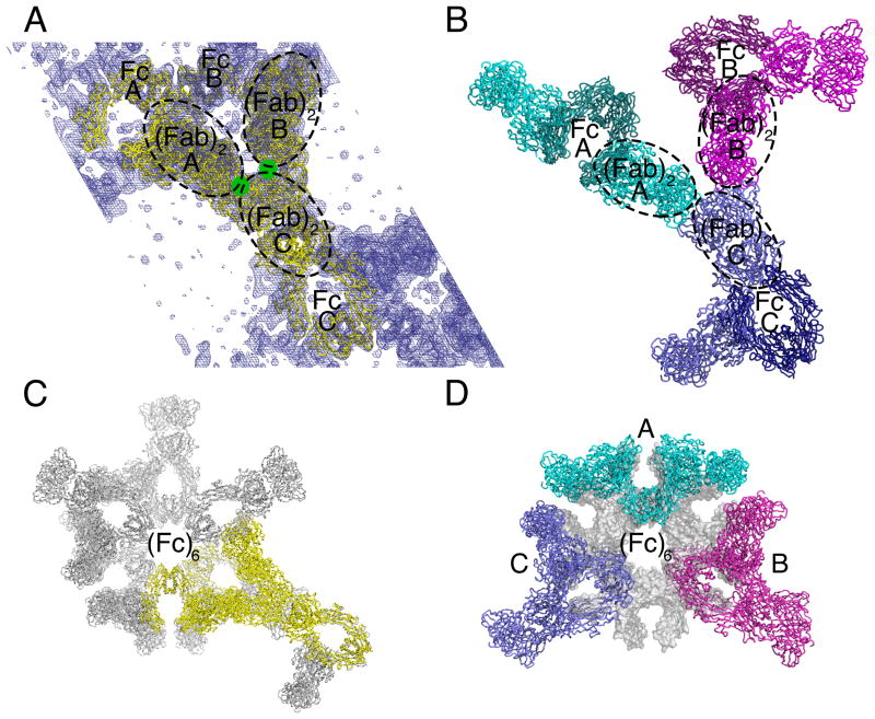
Figure 2. Packing in 2G12 dimer crystals.
(A) Asymmetric unit of a solvent flattened 8.0 Å resolution 2Fo−Fc electron density map contoured at 1.5 σ. The asymmetric unit contained three half-dimers; i.e., three Fc regions and three (Fab)2 units from three distinct 2G12 dimers (A, B, and C). Crystal contacts involved the antigen binding sites in the (Fab)2 units (green circles), see also Figure S2. (B) Application of symmetry operators to generate the second half of each 2G12 dimer. Dimer A (cyan) was structurally distinct from Dimers B (magenta) and C (indigo), which exhibited the same conformation. (C) Non-crystallographic six-fold symmetry axis showing a hexamer of Fc regions coincident with the 61 screw axis along the crystallographic c axis. The asymmetric unit is highlighted in yellow. (D) The three dimers in one layer of the crystal, represented by the cyan, magenta, and indigo dimers A, B, and C (panel B), as they fit into the Fc hexamer ring in panel C, see also Figure S2.

