
Figure 12.
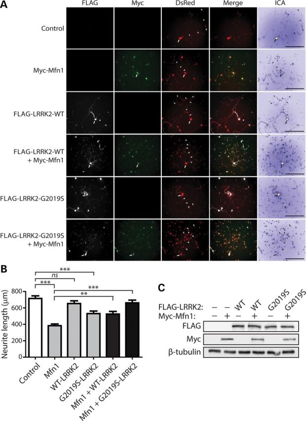
LRRK2 rescues impaired neurite outgrowth induced by Mfn1. (A) Primary cortical neurons were co-transfected with FLAG-tagged LRRK2 (WT or G2019S), Myc-tagged Mfn1 and DsRed-Max constructs at a molar ratio of 10:10:1 at DIV 3 and fixed at DIV 7. Fluorescent microscopic images indicate the co-labeling of cortical neurons with combinations of FLAG-LRRK2, Myc-Mfn1 and DsRed. DsRed images were pseudo-colored with ICA for neurite length measurements. Neuronal soma (arrows) and axonal processes (arrowheads) are indicated. Scale bars: 400 µm. (B) Analysis of DsRed-positive neurites reveals a marked shortening of axonal processes by Mfn1 or G2019S LRRK2 expression alone, with a negligible effect of WT LRRK2 expression alone, compared with control neurites (DsRed alone). Co-expression of G2019S LRRK2 and Mfn1 rescues the Mfn1-induced shortening of axonal processes, whereas WT LRRK2 partially rescues the effects of Mfn1. Bars represent axonal process length (mean ± SEM) expressed as a percent of DsRed alone (control) from ≥90 DsRed-positive neurons taken from at least three-independent experiments/cultures. **P < 0.01 or ***P < 0.001 by one-way ANOVA with Newman–Keuls post hoc analysis. n.s., non-significant. (C) Western blot analysis with anti-FLAG, anti-myc and anti-β-tubulin antibodies of cell extracts derived from rat primary cortical neurons at DIV 7 transiently expressing FLAG-LRRK2 and Myc-Mfn1. The levels of LRRK2 or Mfn1 are not altered when expressed alone or together. Blots are representative of three-independent experiments.