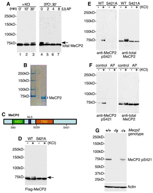
Figure 1. Identification of MeCP2 S421 as a Site of Membrane Depolarization-Induced Phosphorylation.
(A) Antitotal MeCP2 western blots of whole-cell lysates (lanes 1–3) or nuclear extracts treated with alkaline phosphatase (lanes 5– 7) prepared from E18 + 5 DIV dissociated rat cortical neurons harvested at the indicated times following 55 mM KCl stimulation. The arrow indicates the slow-migrating form of MeCP2 induced in response to membrane depolarization. AP: Nuclear extracts were treated with 2, 4, or 8 U of calf intestinal alkaline phosphatase for 30 min at 37°C prior to SDS-PAGE separation. Note: MeCP2 runs at approximately 75 kDa on an 8% SDS-PAGE gel. All protein samples in this study were separated on an 8% gel unless otherwise noted.
(B) Coomassie blue staining of MeCP2 protein purified from rat brain nuclear extract. Both bands were excised and subjected to tandem MS/MS sequencing for identification of phosphorylation sites.
(C) A schematic of MeCP2 illustrating the positions of phosphorylation sites relative to the methyl-CpG-binding domain (MBD), nuclear localization signal (NLS), and the transcriptional repression domain (TRD).
(D) Identification of S421 as a residue required for membrane depolarization-induced phosphorylation of MeCP2. FLAG-tagged wild-type or S421A MeCP2 was transfected into E18 + 2 DIV cortical neurons. Two days later, extracts were prepared from untreated neurons or neurons membrane depolarized for 60 min and subjected to western blotting with the anti-FLAG antibody.
(E) A phospho specific antibody detects phosphorylation of MeCP2 at S421. FLAG immunoprecipitates from E18 + 5 DIV cortical neurons transfected with FLAG-tagged wild-type or S421A MeCP2 were immunoblotted with antitotal MeCP2 or anti-MeCP2 pS421 antibodies. The anti-MeCP2 pS421 antibody recognizes a band at a position corresponding to that of the slow-migrating species of MeCP2.
(F) Membrane depolarization triggers the phosphorylation of MeCP2 at S421. Antitotal MeCP2 and anti-MeCP2 pS421 western blots of nuclear extracts prepared from unstimulated or membrane-depolarized E18 + 5 DIV cortical neurons, either untreated (control) or incubated with alkaline phosphatase for 30 min at 37°C (AP). Unlike other experiments, cultures were not treated with TTX prior to stimulation in this experiment.
(G) The anti-MeCP2 pS421 antibody specifically recognizes MeCP2. Brain lysates from C57BL/6 wild-type Mecp2+/+, heterozygous Mecp2−/+, or Mecp2−/y null mice were probed with anti-MeCP2 pS421 or anti-actin antibodies.

