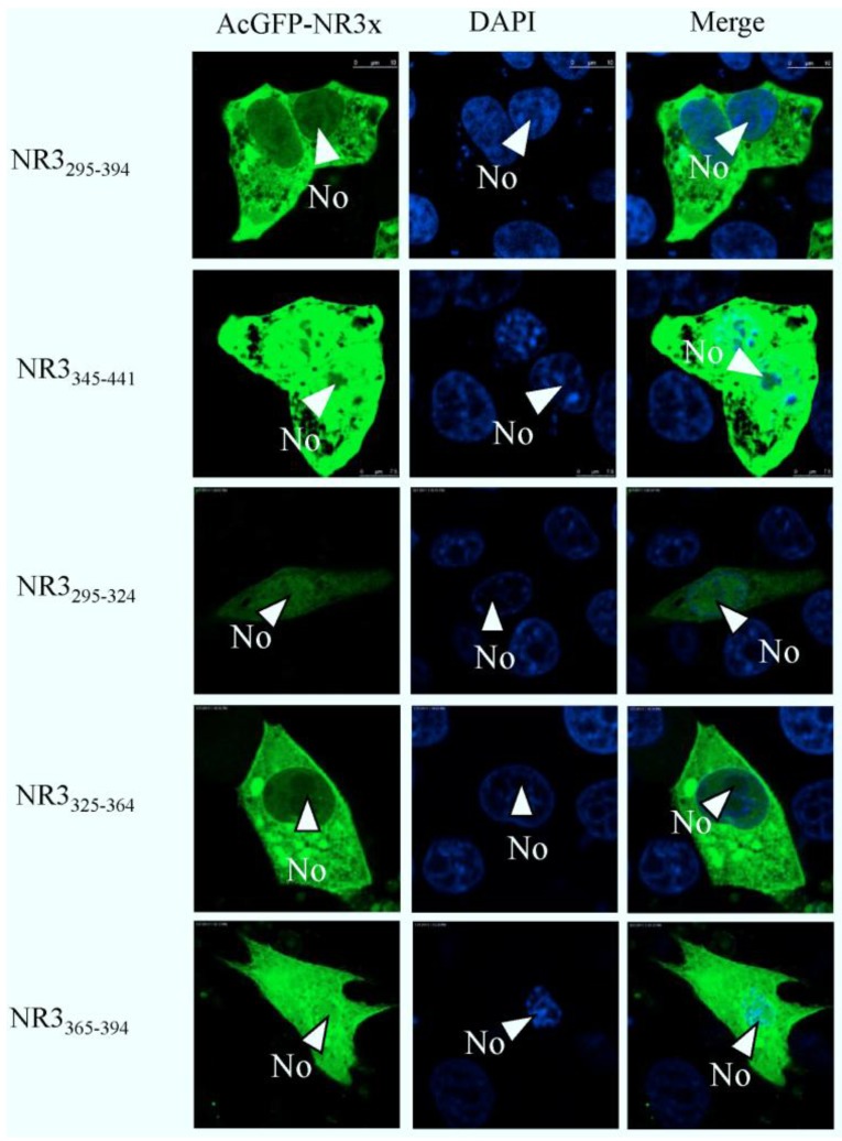
Figure 10.
Confocal microscopy of the sub-cellular localization of fluorescent fusion proteins: AcGFP-N295–394 and AcGFP-N345–441 AcGFP-N295–324, AcGFP-N325–364, AcGFP-N365–394. Fusion proteins are colored green and nucleus colored blue. Merged images are also presented. The nucleolus (No) is arrowed where appropriate.

