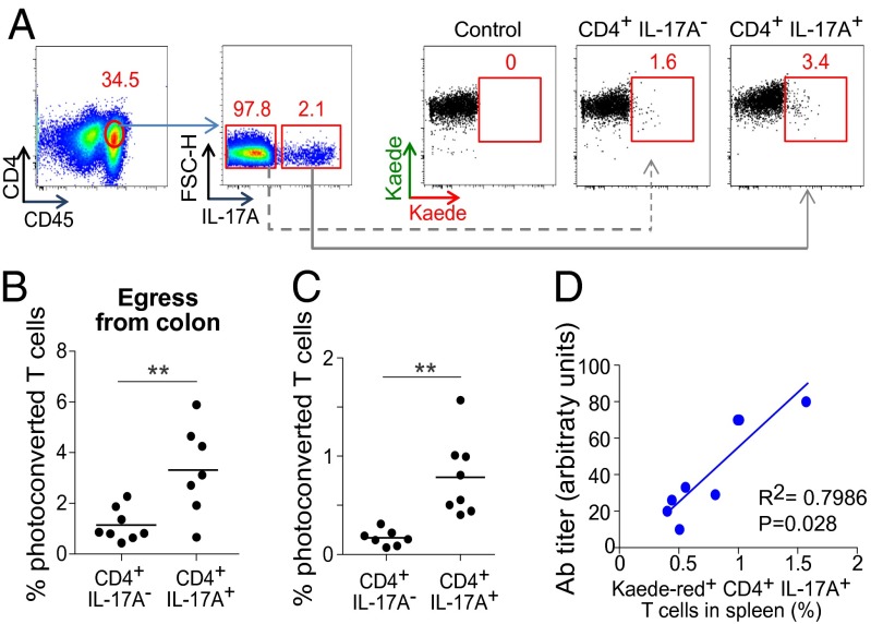
Fig. 4.
Linking colonic T cells to a gut-distal autoinflammatory disease. (A and B) Seventy-two hours after exposure of the descending colon of 24-d-old K/BxN mice to violet light, IL-17A–producing and –nonproducing CD4+ T cells in the PC LI-LP were enumerated, as a confirmation of egress. (A) Representative dot plots; (B) summary data. (C) Immigration to the spleen in these same mice. n = 7–8 from three independent experiments. (D) The proportion of splenic IL-17+CD4+ cells that were Kaede-red+ plotted against the anti-GPI autoAb titer, an early (required) disease indicator in K/BxN mice. **P < 0.01.

