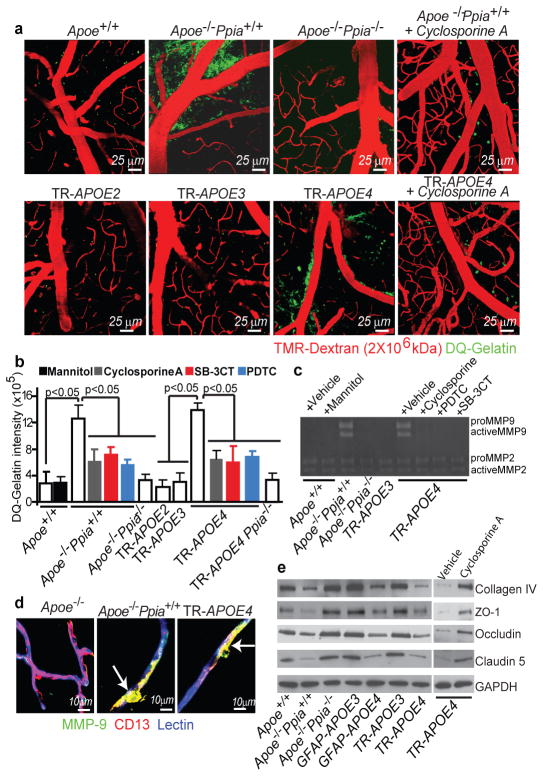
Figure 2. CypA activates NF-kB-MMP-9 pathway causing BBB breakdown in Apoe−/− and APOE4 mice.
(a) Multiphoton microscopy of DQ-gelatin (green) in 8–9-month-old control, Apoe−/− Ppia+/+, Apoe−/− Ppia−/−, TR-APOE2, TR-APOE3, TR-APOE4 and cyclosporine A-treated Apoe−/− Ppia+/+ and TR-APOE4 mice. Red, cortical vessels. (b) Quantification of DQ-gelatin signal in apoE transgenic mice. Effects of cyclosporine A, PDTC, SB-3CT and CypA deletion in Apoe−/− and TR-APOE4 mice. Mean±s.e.m., n=3–6 animals per group.(c) Gelatin zymography of brain tissue in control, Apoe−/− Ppia+/+, Apoe−/− Ppia−/−, TR-APOE3 and TR-APOE4 mice treated with vehicle, cyclosporine A, PDTC or SB-3CT. (d) MMP-9 (green) colocalization with CD13-positive pericytes (red; yellow, merged) in cortical microvessels from 9-month-old Apoe+/+Ppia+/+, Apoe−/− Ppia+/+ and TR-APOE4 mice. Blue, lectin-positive endothelium. Bar=10 mm. (e) Reduced collagen-IV, ZO-1, occludin and claudin-5 levels in 2-week-old Apoe−/− and TR-APOE4 mice and reversal by CypA ablation (Apoe−/− Ppia+/+) and cyclosporine A (TR-APOE4). c and e, representative results from 4–6 experiments.

