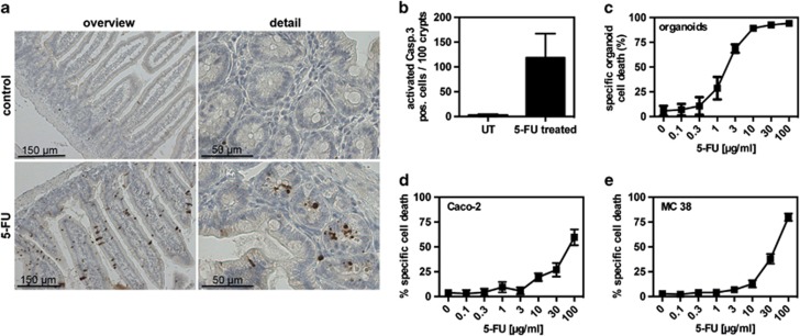
Figure 6.
5-FU induced cell death in vivo, in organoids and in cancer cell lines. (a) Immunohistochemistry of small intestinal tissue sections from control or 5-FU-treated mice (n=3 per group) using an anti-cleaved caspase-3 antibody. Left panel: overview; right panel: detail. (b) Organoids were cultured for 72 h and then treated with different concentrations of 5-FU for 24 h. Cell death was determined by MTT reduction. Data are representative for three independent experiments. (c and d) Caco-2 cells (c) or MC 38 cells (d) were treated with different concentrations of 5-FU for 24 h. Cell death was determined by MTT reduction. Data are representative for three independent experiments

