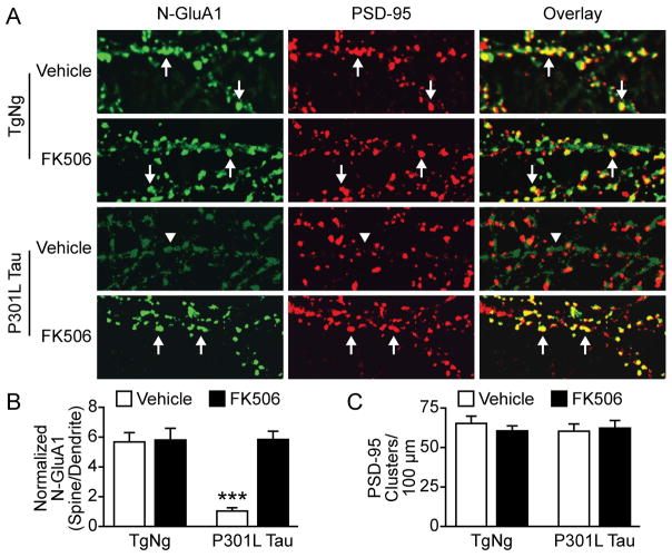
Figure 7.
FK506 rescued the disruption of surface AMPAR clustering caused by P301L tau. (A) Representative images of hippocampal neurons cultured from TgNg mice and transgenic mice expressing P301L tau proteins. The neurons were stained with antibodies to N-GluA1 (green) and PSD-95 (red). At 14–16 DIV neurons were treated with FK506 or DMSO (vehicle) and imaged at 21 DIV. Arrows indicate GluA1 clusters co-localized with PSD-95 clusters; arrowheads indicate diffuse staining of GluA1 in the dendritic shafts. (B) Quantification of N-GluA1 expression in four groups in A. Fluorescent intensity of the green channel was measured at individual dendritic spines and adjacent dendritic shafts. (C) There was no significant difference in the density of spines detected by anti-PSD-95 antibody. Two-way ANOVA, Bonferroni post-test, n=10 neurons per group, ***P < 0.001.

