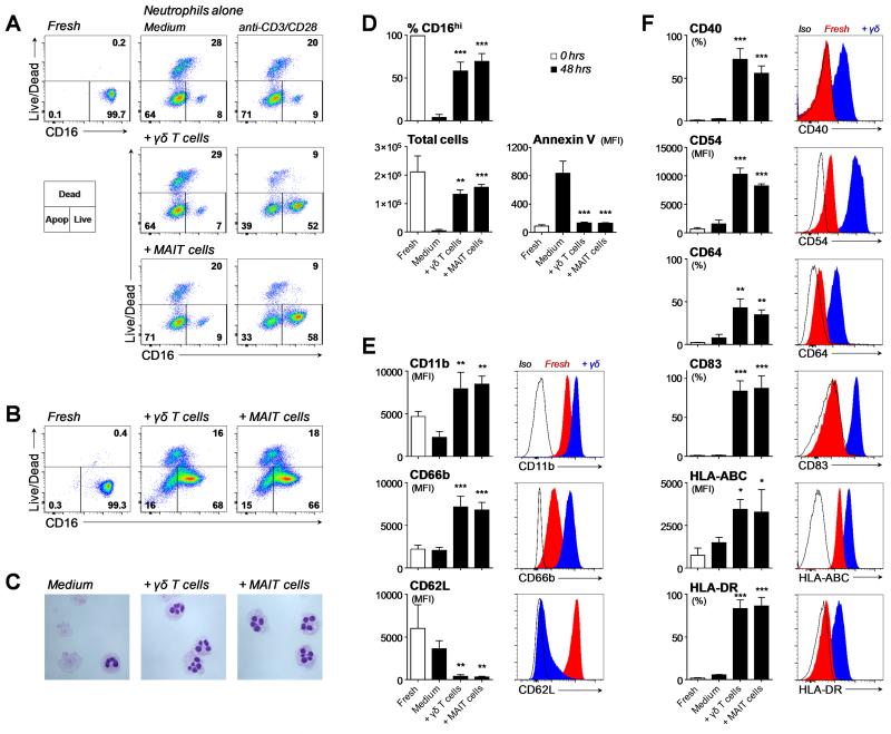
FIGURE 2. Survival, activation and expression of APC markers by unconventional T-cell primed neutrophils.
(A) Neutrophil survival judged by retention of CD16 expression and exclusion of live/dead staining after 48 hour co-culture with FACS-sorted Vγ9/Vδ2 T-cells or MAIT cells, in the absence or presence of anti-CD3/CD28 beads. FACS plots are representative of three donors and depict total neutrophils after gating on CD15+ Vγ9− or CD15+ Vα7.2− cells. (B) Neutrophil survival after 48 hour culture in the presence of HMB-PP activated Vγ9/Vδ2 T-cell or anti-CD3/CD28 activated MAIT cell conditioned medium (representative of three donors). (C) Morphological analysis of surviving neutrophils after 48 hour culture in the absence or presence of HMB-PP activated Vγ9/Vδ2 T-cell or anti-CD3/CD28 activated MAIT cell conditioned medium (representative of two donors). Images were acquired with ×400 original magnification. (D) Neutrophil survival after 48 hour culture in the absence or presence of HMB-PP activated Vγ9/Vδ2 T-cell or anti-CD3/CD28 activated MAIT cell conditioned medium. Shown are means ± SD for the proportion of CD16hi cells (n=9-10), the total number of neutrophils (n=3) and annexin V staining on CD16hi neutrophils (n=3). Expression of (E) activation markers and (F) APC markers on freshly isolated neutrophils and CD16hi neutrophils after 48 hour culture in the absence or presence of HMB-PP activated Vγ9/Vδ2 T-cell or anti-CD3/CD28 activated MAIT cell conditioned medium. Data shown are means ± SD and representative histograms from 3 individual donors. Data were analyzed by one-way ANOVA with Bonferroni’s post-hoc tests; comparisons were made with medium controls. Differences were considered significant as indicated in the figures: *, p<0.05; **, p<0.01; ***, p<0.001.

