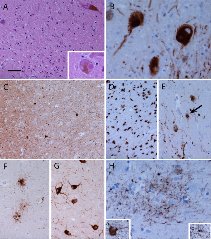
Figure 1.
In Parkinson's disease (PD), there is loss of pigmented neurons from the substantia nigra and remaining neurons may be very sparse (A). Lewy bodies can be observed in residual neurons (A, inset) and are highlighted, together with Lewy neuritis, using α-synuclein immunohistochemistry (B). Lewy bodies and Lewy neurites may be present in significant numbers in the neocortex (C, frontal cortex). In multiple system atrophy (MSA), α-synuclein is primarily deposited in the form of glial cytoplasmic inclusions in oligodendrocytes (D, putamen) and may also form inclusions in neuronal cytoplasm and nuclei (arrow) (E, pontine nuclei). In progressive supranuclear palsy tau forms, aggregates in neurons and glia, giving rise to tufted astrocytes (F, caudate) and neurofibrillary tangles (G, pontine nuclei). A characteristic feature of corticobasal degeneration (CBD) is the astrocytic plaque, formed from aggregated tau in the distal processes of astrocytes (H, parietal cortex). In CBD, tau also accumulates in neurons in the form of neurofibrillary tangles (H, inset a) and in oligodendrocytes as coiled bodies (H, inset b). (A) Haematoxylin and eosin; (B–D) α-synuclein immunohistochemistry; (F–H) tau immunohistochemistry. Bar in (A) represents 100 µm in (C); 50 µm in (A, D–G); 25 µm in inset A, B and H. Pathological images kindly provided by Dr Janice Holton, Queen Square Brain Bank for Neurological Disorders, London.

