
Figure 1. Demonstrating co-registration and segmentation approaches for the analysis of MRI images from the mouse brain.
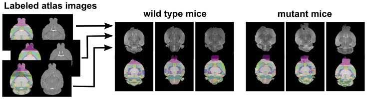
Each of the manually labeled brain was co-registered to each sample's B0 image. The corresponding labels (illustrated as a semi-transparent color overlay) were warped into the sample space and merged via STAPLE. Resulting anatomical labels are illustrated for single wild type and mutant type samples. The olfactory bulb was not included in this analysis due to the variability in extracting the brain tissue leading to incomplete extraction of the olfactory bulbs in some cases. While the resulting labels were probabilistic, the hard segmentations seen here were used for visualization and quality control, and were created by assigning the label of highest probability at each voxel. The color indicates the different regions of the brain.