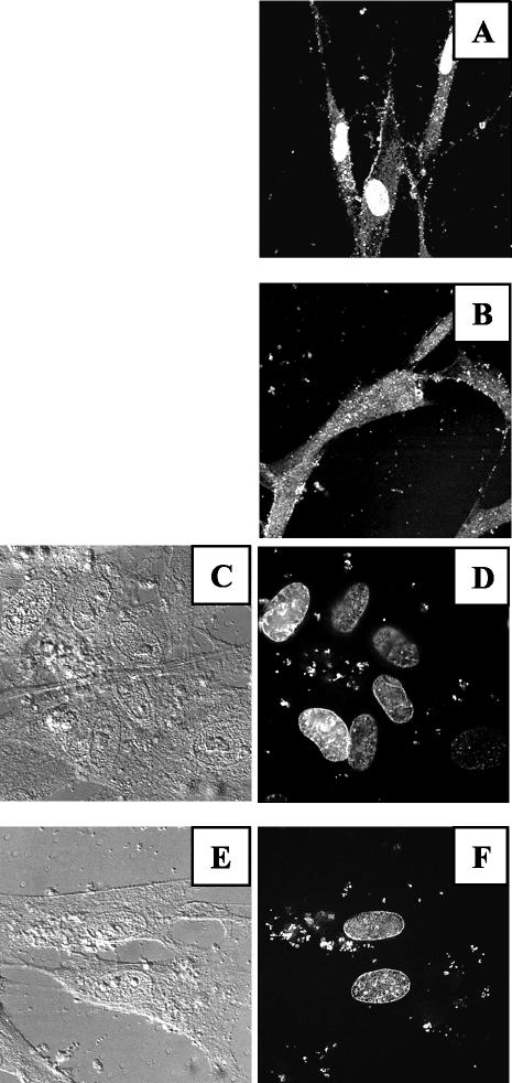
FIG. 8.
The role of cPLA2 in infection is at a postentry step. MRC5 cells were seeded on glass coverslips in six-well plates (2.5 × 10⁵/well), deprived of FCS for 16 h, and incubated for 1 h at 4°C with HCMV (AD169, MOI = 100) untreated (A) or treated (B) with MAFP. Unadsorbed virus was removed with PBS, and cells were further incubated at 37°C for 4 h. Staining was performed with anti-pp65 MAb and observed by confocal microscopy (original magnification, ×60). For panels C to F, MRC5 cells were treated as described for panels A and B, except that DAPI was used to visualize viral DNA. Shown are the phase and DAPI fluorescence pictures for cells infected with untreated HCMV (C and D) or MAFP-treated virus (E and F). Cells were visualized by confocal microscopy (original magnification, ×60).

