
Figure 6.
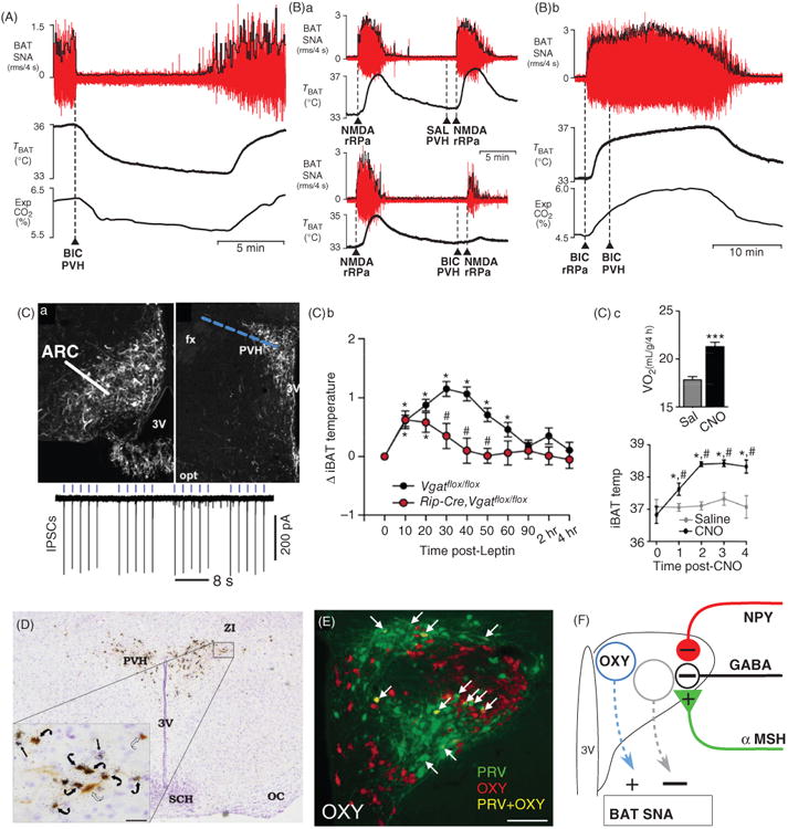
Paraventricular hypothalamic nucleus (PVH) mechanisms influencing BAT thermogenesis. (A) Unilateral nanoinjection of bicuculline (BIC) into the PVH completely reversed the cooling-evoked increases in BAT sympathetic nerve activity (SNA), BAT temperature (Tbat), and expired CO2 (Exp CO2). Modified, with permission, from (197). (Ba) The increases in BAT and Tbat evoked by activation of rRPa neurons with nanoinjection of NMDA are unaffected by unilateral saline injection into the PVH (upper panel), but are markedly reduced by increasing the activity of PVH neurons with a unilateral nanoinjection of bicuculline (BIC) in the PVH (lower panel). Modified from (197). (Bb) Nanoinjection of BIC into the rostral raphe pallidus (rRPa) increases BAT, Tbat, and Exp CO2, and prevents the inhibition of BAT evoked by unilateral nanoinjection of BIC into the PVH. Modified from (197). (Ca) Channelrhodopsin2 (CHR2)-tranfected, RIP-Cre neurons (left panel) and their terminals in PVH (right panel) following adeno-associated virus (AAV) injection in the arcuate nucleus (ARC) of a RIP-Cre mouse; 3V, third ventricle; opt, optic chiasm; fx, fornix. Laser light (blue dashes) pulses depolarizing the CHR2-expressing terminals of ARC RIP-Cre neurons in PVH elicits inhibitory postsynaptic currents (IPSCs, lower panel) in a PVH neuron in a brain slice. (Cb) Leptin increases interscapular BAT (iBAT) temperature in vesicular GABA transporter (Vgat)-floxxed mice, but not in RIP-Cre, Vgatflox/flox mice. (Cc) Following injection into ARC of a cre-dependent AAV producing expression of the designer receptor (hM3Dq) exclusively activated by designer drug (DREADD), selective activation of ARC RIP-Cre neurons with clozapine-N-oxide (CNO) elicits a significant increase in oxygen consumption (VO2) and in iBAT temperature (Temp). Panels Ca-Cc were modified, with permission, from (166). (D) Histological section through the PVH illustrating the overlap of transsynaptically infected, pseudorabies virus (PRV)-labeled neurons (brown) following PRV injections into interscapular BAT and in situ hybridization for melanocortin 4-receptor (MC4-R) mRNA expression (black granules). Inset: High magnification of the outlined portion of the PVH. Note the presence of PRV in neurons surrounded by MC4-R (curved black arrows) and PRV in neurons without associated MC4-R (curved open arrows). Modified, with permission, from (349); 3V, third ventricle; ZI, zona inserta; oc, optic chiasm; SCH, suprachiasmatic nucleus. E: immunohistochemical labeling of PVH neurons for oxytocin (OXY, red) and for transsynaptic infection with PRV (green) after PRV injections into interscapular BAT. Arrows indicate neurons containing both PRV and OXY (yellow). Modified, with permission, from (259). (E) Schematic, based in part on (71), of the local PVH neurocircuitry proposed to mediate the GABAergic, neuropeptide Y (NPY) and α-melanocyte-stimulating hormone (αMSH) influences on BAT thermogenesis mediated by PVH neurons.