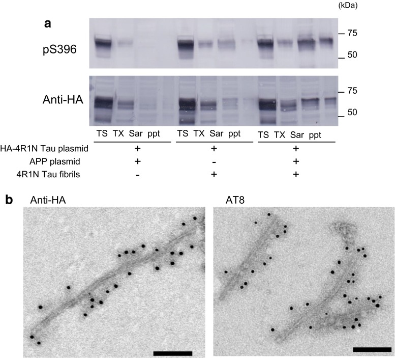
Fig. 4.
Immunoblotting and immunoelectron microscopic analyses of tau in cells expressing both APP and HA-tagged 4R1N tau. Immunoblot analysis of lysates from cells transfected with both HA-4R1N tau and APP, cells transfected with HA-4R1N tau and treated with 4R1N tau fibrils, and cells transfected with both HA-4R1N tau and APP and treated with 4R1N tau fibrils (a). Cells were sequentially extracted to obtain Tris-soluble (TS), Triton X-100-soluble (TX), and Sarkosyl-soluble (Sar) fractions, leaving the pellet fraction (ppt). HA-4R1N tau was detected with pS396 (p-Ser-396) and anti-HA antibodies. Immunoelectron microscopy of tau in the Sar-insoluble fraction from cells transfected with both HA-4R1N tau and APP and treated with 4R1N tau fibrils (b). Anti-HA-positive (left panel) and AT8 (p-Ser-202 and p-Thr-205)-positive (right panel) filaments were observed. Scale bars represent 100 nm

