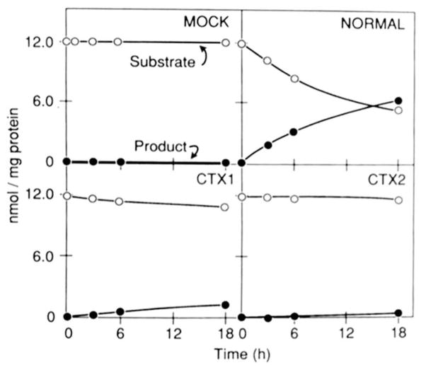
Fig. 4. Expression of normal and CTX sterol 27-hydroxylase cDNAs.
COS cells were transfected with the indicated cDNA in the pCMV2 vector. Forty-eight hours after transfection, the medium was made 2.5 μM in substrate (5β-[7β-3H]cholestane-3α,7α,12α-triol) as outlined under “Experimental Procedures” and left on the cells for the indicated time periods. The conversion of substrate into products (5β-[7β-3H]cholestane-3α,7α,12α,27-tetrol plus 3α,7α,12α-trihydroxy-5β-[7β-3H]cholestanoic acid) was determined by thin layer chromatography analysis of media sterols. The results shown are representative of two separate transfection experiments.

