
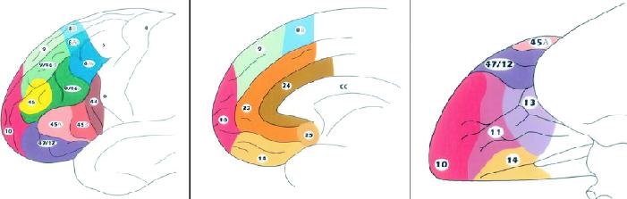
Fig. 2: Schematic diagrams of the lateral (left), medial (middle) and inferior (right) surfaces of the human frontal lobe to illustrate its cytoarchitectonic parcellation. Reproduced with permission from Elsevier.74
Official websites use .gov
A
.gov website belongs to an official
government organization in the United States.
Secure .gov websites use HTTPS
A lock (
) or https:// means you've safely
connected to the .gov website. Share sensitive
information only on official, secure websites.
Fig. 2: Schematic diagrams of the lateral (left), medial (middle) and inferior (right) surfaces of the human frontal lobe to illustrate its cytoarchitectonic parcellation. Reproduced with permission from Elsevier.74